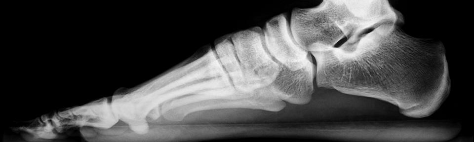
Tumor Pathology- Bone Lesion
When you identify a bone lesion, follow this basic checklist to help you accurately describe the lesion and narrow your differential diagnosis:
Bone Tumors and Tumorlike Conditions: Analysis with Conventional Radiography
Theodore T. Miller
Radiology 2008 246:3, 662-674
A Modified Lodwick-Madewell Grading System for the Evaluation of Lytic Bone Lesions
Jamie T. Caracciolo, H. Thomas Temple, G. Douglas Letson, and Mark J. Kransdorf
American Journal of Roentgenology 2016 207:1, 150-156
- Patient demographics
- Age
- Medical history
- Clinical concern for infection?
- Known cancer?
- Systemic or metabolic disease?
- Location
- Bone involved
- Axial vs appendicular skeleton
- Flat vs long bones
- Location within the bone
- Longitudinal position
- Epiphysis, metaphysis or diaphysis
- Transverse position
- Intramedullary (central or eccentric)
- Cortical
- Juxtacortical
- Longitudinal position
- Number
- Single vs Multiple
- Bone involved
- Morphologic features
- Margins
- Periosteal reaction
- Matrix calcifications

Many bone lesions occur in characteristic locations with predilection for certain age groups.
Peak age predelection bone lesions at <20 years old:
Malignant:
- Leukemia
- Ewing sarcoma
- Osteosarcoma (conventional, periosteal, telangiectatic)
- Rare metastases (retinoblastoma, neuroblastoma)
- Rhabdomyosarcoma
- Hodgkin lymphoma
Benign:
- Fibrous cortical defect
- Nonossifying fibroma
- Simple bone cyst
- Aneurysmal bone cyst
- Chondroblastoma
- Langerhans cell histiocytosis
- Osteoblastoma
- Osteofibrous dysplasia
- Fibrous dysplasia
- Enchondroma

Peak age predelection bone lesions at 20-40 years old:
Malignant:
- Osteosarcoma (parosteal)
- Adamantinoma
Benign:
- Giant cell tumor
- Enchondroma
- Osteoblastoma
- Osteoid osteoma
- Chondromyxoid fibroma
- Fibrous dysplasia

Peak age predelection bone lesions at >40 years old:
Malignant:
- Metastatic disease
- Myeloma
- Chondrosarcoma
- Osteosarcoma (secondary)
- Non-Hodgkin lymphoma
- Undifferentiated pleomorphic sarcoma
Benign:
- Paget disease
- Fibrous dysplasia
The terminology used in modified Lodwick classification is useful for describing morphologic features with higher grades reflecting increasingly aggressive features and higher risk that findings represent malignancy.
Trace lesion margins closely use the following descriptors to help characterize margins:
- Geographic, well-defined, sharp margin with thin rim of sclerosis (Grade IA, Fig A)
- Geographic, well defined sharp margin with clear distinction between lucency and normal bone (grade 1B, Fig B)
- Geographic, Ill-defined zone of transition (Grade II, Fig E)
- Focal change in margin or progressive endosteal scalloping (Grade IIIA)
- Moth eaten or permeative osteolysis- scattered, confluent holes in the cortex that fade into normal bone with wide zone of transition (Grade IIIB, Fig F)
Note that lesions with geographic features may still represent malignant process (I.e. plasmacytoma, metastases) while lesions with aggressive features may represent a benign process (ex. Osteomyelitis or LCH).
Lesions without pathognomoic features often require further workup with MRI and/or biopsy for definitive diagnosis.

Fig A. Geographic, well-defined lucent lesion with thin rim of sclerosis in the calcaneus with location and characteristic findings of intraosseous lipoma.

Fig C. Geographic, well-defined lucent lesion with narrow zone of transition. Features are most consistent with non-ossifying fibroma of the distal tibia.

Fig E. Geographic, lytic lesion with wide zone of transition centered within anterior cortex of mid tibial diaphysis. Given history of lung cancer, this is favored to represent metastatic disease.

Fig B. Geographic, slightly expansile lucent lesion within left iliac wing with differential considerations including both benign and malignant etiologies such as aneursymal bone cyst, plasmacytoma, or metastatic disease. Consider further characterization with MRI as clinically indicated.

Fig D. Geographic, well-defined expansile lesion with narrow zone of transition. Aneurysmal bone cyst of the distal radius.

Fig F. Permeative, lytic lesion within mid humeral diaphysis with wide zone of transition and areas of cortical disruption, consistent with aggressive lesion. Recommend MRI for further assessment.
Some lesions demonstrate associated periosteal reaction, the pattern of which can be helpful for determining growth rate and aggressiveness.
- Solid periosteal reaction- nonaggressive, slow growing process. (Fig A)
- Lamellated periosteal reaction- aggressive, waxing and waning growth. (Fig B)
- Spiculated/sunburst periosteal reaction- aggressive, rapid growth. (Fig C)
- Codman triangle- aggressive, often subtle lifting of the periosteum away from cortex suggesting rapid growth. (Fig D)

Fig a. Eccentric, cortically based lucency with surrounding smooth periosteal reaction in mid posterior tibial diaphysis, possibly represents osteoid osteoma. Consider CT for further characterization.

Fig c. Permeative lytic lesion within proximal humeral metadiaphysis with surrounding aggressive sunburst periosteal reaction.

Fig b. Lytic lesion centered within lateral femoral metaphysis with Ill-defined margins and aggressive features, including cortical breakthrough and lamellated periosteal reaction.

Fig d. Permeative lytic lesion within mid humeral diaphysis with aggressive periosteal lifting at both proximal and distal lateral cortex.
Lesions may appear lytic, sclerotic, or mixed, demonstrating a specific pattern of internal matrix calcification, that can help classify the lesion.
- Osteoid matrix- fluffy, amorphus, cloudlike mineralization, which may extend beyond native cortex if aggressive. (Fig A, Fig B)
- Chondroid matrix- punctate, ring, and arc-like mineralization. (Fig C, Fig D)
- Ground glass matrix- subtle, homogenous and faint mineralization, often seen in benign fibro-osseous lesions. (Fig E)

Fig A. Mixed lytic and sclerotic lesion of proximal humerus with fluffy, osteoid matrix extending beyond the cortex medially and aggressive periosteal reaction, concerning for osteosarcoma.

Fig C. Small focus of ring and arc calcifications within proximal humeral metaphysis without aggressive features, favored to represent low grade chondroid lesion such as enchondroma.

Fig E. Intramedullary lesion within femoral metadiaphysis with ground glass internal matrix with differential considerations including non-ossifying fibroma or simple bone cyst.

Fig B. Mixed lytic and sclerotic lesion with extensive sunburst periosteal reaction. Features concerning for osteosarcoma of the left proximal humerus.

Fig D. Lesion of the left distal femoral metaphysis with ring and arc matrix and extra-osseous/soft tissue component. Features are concerning for chondrosarcoma.
Lesions with or without radiographically apparent aggressive features may feature a soft tissue component often appearing as abnormal effacement of fat planes surrounding the lesion. This is highly suggestive of malignant etiology and is often seen it the setting of osteosarcoma, Ewing sarcoma, and lymphoma.

Large permeative lesion with wide transition zone demonstrates cortical breakthrough and extensive surrounding soft tissue component, consistent with aggressive malignant lesion.
When evaluating any fracture, be sure to assess of underlying pathologic lesion (Fig A). Likewise, if a bone lesion is known look closely for any signs of acute pathologic fracture (Fig B).

Fig A. Mildly displaced spiral fracture of distal femoral diaphysis with underlying permeative, lytic lesion, consistent with pathologic fracture.

Fig B. Minimally displaced pathologic fracture of proximal humeral cortex at the lateral margin of expansile, lytic lesion demonstrating features consistent with aneurysmal bone cyst.