
We have developed a comprehensive diffeomorphic registration process allows accurate MRgFUS treatment evaluation that accurately maps tissue structure between treatment and post treatment MRI images and through all steps of processing to the final histopathology. This comprehensive registration process enables development of accurate histology-verified cell viability treatment biomarkers. We have also developed and rigorously tested an in vivo MRI to volumetric histopathology registration pipeline that estimates and corrects for deformation at each tissue processing step, accounting for orientation loss and shrinkage from excision and fixation, loss of morphology from gross slicing, and shearing and stretching from microscopic sectioning. This landmark-free pipeline achieves sub-voxel registration accuracy (<1 mm) between in vivo MRI and volumetric histopathology, a 30-77% improvement over the target registration error of previous methods, even compared to methods that use landmarks-driven registration or only evaluate select 2D slices.
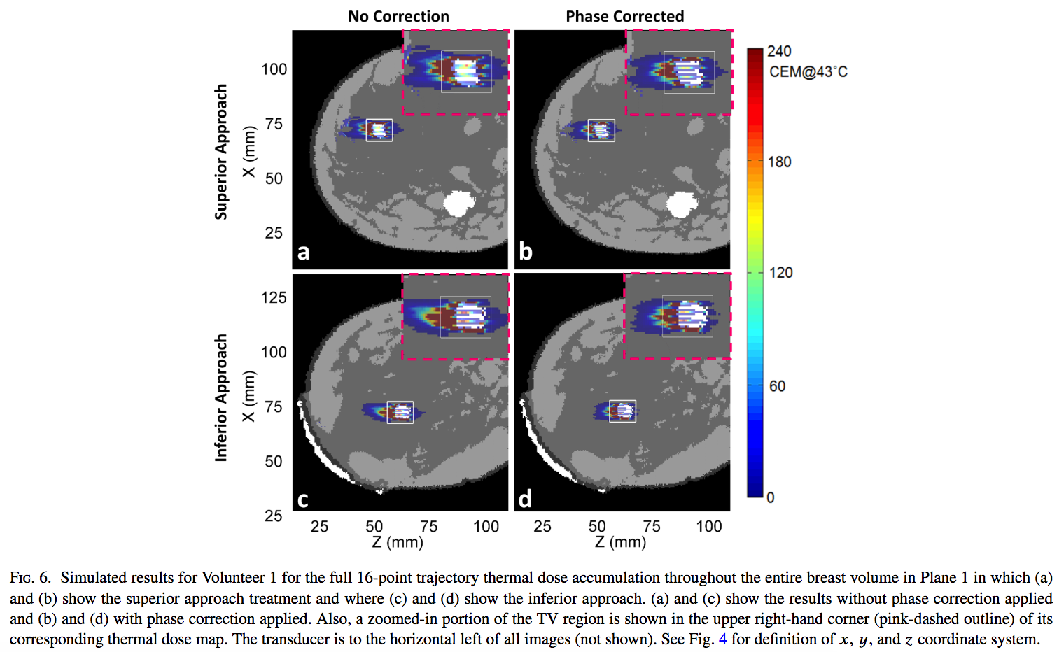
The ability to accurately and reliably model ultrasound beam propagation is a crucial component of MRgFUS research. Modeling patient-specific treatments allows us to examine potential phase aberration effects and ways to correct for those effects, all before actually using the technology in an experiment or with a patient. Accurate acoustic modeling can result in safer and more effective treatments. It is important to be able to accurately model how a focused ultrasound beam propagates through the inhomogeneous tissue structures of the human body. Our lab group has developed a technique known as the hybrid angular spectrum (HAS) technique, which is an extension of the more traditionally used angular spectrum method. HAS can successfully model linear wave propagation and can account for the inhomogeneous structure of human tissues. Our current research is quantitatively validating our acoustic simulation method in ex vivo and in vivo environments and will be integrated into Thermoguide, a MRgFUS clinical treatment environment developed by Image Guided Therapy, Inc.
Related Publications
Almquist, S., Parker, D. L., & Christensen, D. A. (2016). Rapid full-wave phase aberration correction method for transcranial high-intensity focused ultrasound therapies. J Ther Ultrasound, 4(1), 30. doi:10.1186/s40349-016-0074-7
Farrer, A. I., Almquist, S., Dillon, C. R., Neumayer, L. A., Parker, D. L., Christensen, D. A., & Payne, A. (2016). Phase aberration simulation study of MRgFUS breast treatments. Medical physics, 43(3), 1374. doi:10.1118/1.494101
Dillon, C. R., Farrer, A., McLean, H., Almquist, S., Christensen, D., & Payne, A. (2018). Experimental assessment of phase aberration correction for breast MRgFUS therapy. Int J Hyperthermia, 34(6), 731-743. doi:10.1080/02656736.2017.142202
Vyas, U., & Christensen, D. (2012). Ultrasound beam simulations in inhomogeneous tissue geometries using the hybrid angular spectrum method. IEEE Trans Ultrason Ferroelectr Freq Control, 59. doi:10.1109/tuffc.2012.230
Johnson, S. L., Dillon, C., Odeen, H., Parker, D., Christensen, D., & Payne, A. (2016). Development and validation of a MRgHIFU non-invasive tissue acoustic property estimation technique. Int J Hyperthermia, 32(7), 723-734. doi:10.1080/02656736.2016.121618
Vyas, U., & Christensen, D. A. (2011). Extension of the angular spectrum method to calculate pressure from a spherically curved acoustic source. J Acoust Soc Am, 130(5), 2687-2693. doi:10.1121/1.362171
Zimmerman BE, Johnson SL, Odéen HA, Shea JE, Factor RE, Joshi SC, Payne A (2021). Histology to 3D in vivo MR registration for volumetric evaluation of MRgFUS treatment assessment biomarkers. Sci Rep, 11(1), 18923.
Zimmerman B, Johnson S, Odéen H, Shea J, Foote MD, Winkler N, Joshi S, Payne A (2021). Learning multiparametric biomarkers for assessing MR-guided focused ultrasound treatments. IEEE Trans Biomed Eng, 68(5):1737-1747.