Traumatic Foot Injury

Foot radiographs obtained after trauma should evaluate for fracture, dislocation and signs of ligamentous injury.
Familiarize yourself with common locations and patterns of injury involving the digits, metatarsals, midfoot, and hindfoot.

Great toe distal phalanx fracture
- Most common fracture in the foot
Sesamoid fracture
- Hyperextension injury or dorsal dislocation of the first metatarsophalangeal joint
- Associated with plantar plate injury
Bipartite sesamoid
- Normal variant often confused for fracture
- Distinguished by well corticated margins with the sum of two parts being larger that the adjacent normal hallux sesamoid.

Lisfranc fracture-dislocation
- The Lisfranc ligament connects the medial cuneiform to the base of the 2nd metatarsal and is important for stabilizing the arch of the foot.
- Disruption will present as subluxations or dislocations of the tarsometatarsal joints, widening at the Lisfranc interval, and/or ossific flecks (avulsion fragments) at the Lisfranc interval.
- Injury can be difficult to detect.
- Weight bearing radiographs can help highlight widening at the interval
- Contralateral foot radiographs are helpful in the evaluation of symmetry
- With high enough clinical suspicion, cross sectional imaging may be needed.
- CT or weightbearing CT- avulsion fracture, malalignment
- MRI- ligamentous complex injury
FINDINGS:
Moderate lateral subluxation of the second through fourth tarsometatarsal joints. Mildly displaced, intraarticular fractures of the second through fourth metatarsal bases and lateral aspect of lateral cuneiform with comminuted, displaced fragments within first intermetatarsal space.

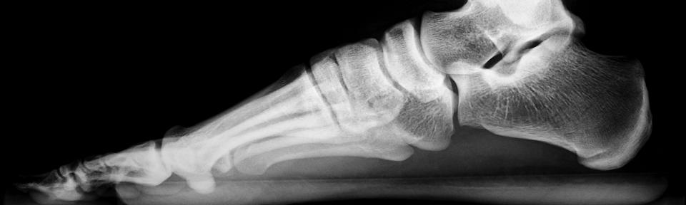
Fig 1- Initial radiograph. No visible acute fracture.

Fig 2- 8 week follow up radiograph. Nondisplaced second and third metacarpal fractures with associated periosteal reaction and callus formation.
Metatarsal stress fracture
- Thin, cortical lucency with associated stress reaction
- Often not radiographically apparent until cortical thickening and/or periosteal reaction has formed. (Fig. 2)
- If clinical concern, MRI may be recommended.


Fifth metatarsal fractures
- Avulsion of the 5th metatarsal styloid
- aka “pseudo-Jones” aka “dancer fracture”
- Insertion of the peroneus brevis
- Jones fracture
- Transverse fracture of proximal 5th metatarsal at the level of the 4th and 5th intermetatarsal articulation
- Risk of non-union
- Diaphyseal fracture
- Usually stress fracture
1st FINDINGS:
Minimally displaced avulsion fracture of the base of the 5th metatarsal
2nd FINDINGS:
Nondisplaced transversely oriented fracture through the proximal fifth metatarsal with extension to intermetatarsal joint (Jones Fracture).
3rd FINDINGS:
No changes.

Example report negative for traumatic injury
FINDINGS:
- No acute fracture or dislocation. Joint spaces are preserved. Midfoot alignment is intact. (only if weightbearing)
IMPRESSION:
- No acute fracture or traumatic malalignment.
- Evaluation of the Tarsometatarsal Joint Using Conventional Radiography, CT, and MR Imaging. Nasir A. Siddiqui, Mauricio S. Galizia, Emad Almusa, and Imran M. Omar. RadioGraphics 2014 34:2, 514-531
- Fractures of Proximal Portion of Fifth Metatarsal Bone: Anatomic and Imaging Evidence of a Pathogenesis of Avulsion of the Plantar Aponeurosis and the Short Peroneal Muscle Tendon. Daphne J. Theodorou, Stavroula J. Theodorou, Yousuke Kakitsubata, Michael J. Botte, and Donald Resnick. Radiology 2003 226:3, 857-865.