Biochemistry Research Advances

Biochemistry Research Advances
2025
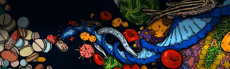
How did our antiviral immune system evolve?
Hundreds of millions of years ago, animals used a process called RNA interference (RNAi) to fight viruses. During evolution, viruses and their animal hosts performed an intricate dance of defense and counter defense. Viruses acquired mutations or new functions to escape destruction by RNAi, and animals retaliated with their own changes to maintain robust antiviral RNAi; this process occurred over and over. To understand the details of how antiviral defense evolved, we used a process called Ancestral Protein Reconstruction to predict the sequence of the ancestral helicases that enabled RNAi. This allowed us to synthesize ancient helicases and test their activities in the laboratory. We found that the most ancient helicase (AncD1D2) bound viral dsRNA tightly and also used ATP hydrolysis to fuel processive movement, or translocation (arrow), along the dsRNA to enable its destruction. As RNAi helicases evolved, dsRNA binding, ATP hydrolysis, and translocation activities declined until they were completely lost in the vertebrate ancestor that gave rise to the modern human helicase. After a brave fight, the RNAi helicase gave up, and a whole new antiviral pathway took over. The vestigial helicase in modern humans adopted a new role in processing small RNAs called microRNAs.
Aderounmu AM, Maus-Conn J, Consalvo CD, Shen PS, Bass BL
Proc Natl Acad Sci, 2025 Jun 3;122(22).

A new drug lead for chronic pain from a fish-hunting cone snail
Persistent pain affects one in five people worldwide, often with severely debilitating consequences. While current treatments can be effective for mild or acute pain, they are largely inadequate for managing moderate to severe chronic pain, underscoring the urgent need for new therapeutics. In a new study, the Safavi lab revealed that certain species of fish-hunting cone snails have evolved a venom-derived strategy that potently and selectively targets the somatostatin receptor 4 (SSTR4), a receptor important for pain signaling. Mining the venom of hundreds of cone snails, the authors identified consomatin Fj1, the most potent and selective peptide agonist of the SSTR4 known to date. Peripheral administration of synthetic consomatin Fj1 produced robust pain reduction in mouse models of postoperative and neuropathic pain. Structure-activity-guided optimization further yielded Fj1 analogs with enhanced potency and selectivity. Together, these findings demonstrate how venom-derived peptides can exploit peripheral signaling for pain modulation and highlight cone snail venoms as a rich source of new drug leads for the treatment of persistent pain. This research formed the foundation for a patent application and the creation of a biotechnology start-up dedicated to translating this novel pain therapeutic into the clinic, with the potential to significantly improve outcomes for patients with chronic pain.
TL, Engholm E, Yeung HY, Sørensen KK, Goddard CM, Jensen KL, Smith NA, Martin LF, Smith BJ, Madsen KL, Jensen KJ, Patwardhan A, Safavi-Hemami H.
Sci Rep. 2025 Nov 28;15(1):42638.

Why do heme crystals wildly spin inside malaria parasites that infect red blood cells?
Malaria is an ancient scourge of humanity caused by single-cell Plasmodium parasites that infect and grow inside human red blood cells. This disease still kills over 600,000 people each year, primarily in Africa. During infection, parasites consume and digest the abundant hemoglobin protein. This process releases lots of the oxygen-carrying cofactor, heme, which the parasite packages into dense heme crystals. Forming these crystals is essential for parasite survival and a major antimalarial drug target. These crystals rapidly spin around inside parasites, but the nature and significance of this motion have been mysterious. This study revealed that these crystals trigger the breakdown of hydrogen peroxide on their surface, which provides an energetic “thrust” that propels crystals to dance around inside parasites. Hydrogen peroxide, which is toxic to the parasite, is produced as a byproduct of digesting hemoglobin. The spinning crystals help to “burn off” excess peroxide before it can harm the parasite. Overcoming this defense mechanism could lead to better antimalarial treatments. These results also reveal how nanoparticles move in confined spaces inside cells, which can help engineers design new biology-inspired propulsion and drug-delivery systems at tiny scales.
Chemical propulsion of hemozoin crystal motion in malaria parasites
Hastings EM, Skóra T, Carney KR, Fu HC, Bidone TC, Sigala PA.
Proc Natl Acad Sci U S A. 2025 Nov 4;122(44). Also highlighted in @THEU and Price Engineering

2024
Caenorhabditis elegans Dicer acts with the RIG-I-like helicase DRH-1 and RDE-4 to cleave dsRNA
All organisms have pathways to fight viruses. Vertebrates, including humans, use proteins called RIG-I-like receptors to detect viral double-stranded RNA (dsRNA) and induce an interferon immune response. Invertebrates do not have interferon pathways and instead use RNA interference (RNAi), which involves cleavage of viral dsRNA by an enzyme called Dicer. Studies in the invertebrate worm C. elegans led to the Nobel prize-winning discovery of RNAi, yet, more than two decades after this discovery, C. elegans Dicer remained biochemically uncharacterized. In this paper we overcome challenges to purification of C. elegans Dicer by co-expression with its protein partners, a dsRNA binding protein called RDE-4, and surprisingly, a protein that looks just like the RIG-I like receptor used in human antiviral defense. Using biochemical assays, as well as cryo-electron microscopy to reveal three-dimensional shape, we determined that the C. elegans antiviral complex exhibits properties found in other antiviral Dicers, but has parsed the activities often intrinsic to Dicer, to other proteins in the antiviral complex. Most notably, it is DRH-1 that is primarily responsible for ATP hydrolysis and threading of dsRNA, rather than Dicer.
Caenorhabditis elegans Dicer acts with the RIG-I-like helicase DRH-1 and RDE-4 to cleave dsRNA
Consalvo CD, Aderounmu AM, Donelick HM, Aruscavage PJ, Eckert DM, Shen PS, and Bass BL.
eLife, 2024. 13:RP93979.

Structural basis for blood pressure drug function
Thiazide (e.g., hydrochlorothiazide) and thiazide-like diuretic drugs (e.g., chlorthalidone and indapamide) have been essential medications for lowering blood pressure and for treating fluid retention since the 1950s. They do so by acting on and preventing the Na+-Cl- ion co-transporter (NCC) from absorbing salt (and water) in the kidneys. Our NCC structures caught hydrochlorothiazide, chlorthalidone, and indapamide in the acts of inhibiting NCC from transporting ions. ‘Seeing’ how these drugs work in atomic detail paves the way for future medicinal chemistry efforts to further improve the potency and specificity of this important class of diuretic drugs. Aberrant activation of NCC by kinases, enzymes that add phosphate groups to a key motif of the transporter, cause hypertension. Our kinases activated NCC structures now show how these enzymes change NCC structure and accelerate its ion translocation rate. Our structures imply that small molecule compounds that interfere with this kinase activation process could serve as leads for the development of novel diuretic drugs.
Zhao Y, Schubert H, Blakely A, Forbush B, Smith MD, Rinehart J, Cao E. Structural Bases for Na+-Cl- Cotransporter Inhibition by Thiazide Diuretic Drugs and Activation by Kinases.
Nat Commun. 2024 Aug 14;15(1):7006.

Using templating to accelerate chemical protein synthesis
Chemical protein synthesis (CPS) is an emerging technology that enables precise atomic control of a protein’s composition, enabling the production of custom proteins with non-natural amino acids and complex modifications that are not accessible using standard recombinant techniques. To make a synthetic protein, it is divided into ~50 amino acid segments (produced using standard peptide chemistry), which are then ligated together one-at-a-time to slowly assemble a full-length protein. These ligation reactions are complex and inefficient, requiring many reaction steps and purifications that often result in unusable yields. In this work, we describe a templated ligation technique that allows us to ligate three peptides in a single step with no intermediate purifications to facilitate small protein production. “Templating” is a process that brings each peptide into close proximity with its neighbor, speeding the ligation reaction and improving overall yields. This technology, called CAPTN - Controlled Activation of Peptides for Templated Native Chemical Ligations – greatly accelerates the production of synthetic proteins while minimizing side-reactions, with wide applications in drug discovery, mechanistic studies, and structural biology. Future research will focus on extending this technology to a larger number of peptide segments to enable rapid production of larger proteins, especially valuable for production of mirror-image proteins (composed of D-amino acids) used in mirror-image drug discovery.
Selective Activation of Peptide-Thioester Precursors for Templated Native Chemical Ligations
Spaltenstein P, Giesler RJ, Scherer SR, Erickson PW, Kay MS.
Angew Chem Int Ed Engl. 2025 Jan 2;64(1):e202413644. Epub 2024 Oct 25.

Fish-hunting cone snails use hormone mimics to disrupt blood glucose in their prey
Venomous animals have evolved diverse molecular mechanisms to incapacitate prey and defend against predators. Most venom components disrupt nervous, locomotor, and cardiovascular systems or cause tissue damage. The previous discovery that certain fish-hunting cone snails use weaponized insulins to induce hypoglycemic shock in prey highlighted a unique example of toxins targeting glucose homeostasis. In a new study, Yeung, Safavi-Hemami and colleagues show that, in addition to insulins, the deadly fish hunter, Conus geographus, uses a selective somatostatin receptor 2 (SSTR2) agonist that blocks the release of the insulin-counteracting hormone glucagon, thereby exacerbating insulin-induced hypoglycemia in prey. The native toxin, Consomatin nG1, contains a minimized vertebrate somatostatin-like core motif connected to a heavily glycosylated N-terminal tail. Remarkably, the toxin’s N-terminal tail closely mimics a somatostatin isoform that is specifically found in fish pancreas. Collectively, this study provides a stunning example of chemical mimicry in nature, highlights the combinatorial nature of venom components, and establishes glucose homeostasis as an effective target for prey capture.
Yeung HY, Ramiro IBL, Andersen DB, Koch TL, Hamilton A, Bjørn-Yoshimoto WE, Espino S, Vakhrushev SY, Pedersen KB, de Haan N, Hipgrave Ederveen AL, Olivera BM, Knudsen JG, Bräuner-Osborne H, Schjoldager KT, Holst JJ, Safavi-Hemami H.
Nat Commun. 2024 Aug 20;15(1):6408.
Also highlighted in ScienMag Morning News, Genetic Engineering and Biotechnology News, Cosmos Magazine, and Science Daily

A promising broad-acting SARS-CoV-2 monoclonal antibody
Monoclonal antibodies (mAb) have proven effective and versatile tools for the treatment and prevention of viral infections, including COVID-19. Five mAb therapeutics against SARS-CoV-2 were authorized for clinical use in the first several years of the pandemic and saw widespread utility, but each lost activity soon after authorization due to the evolution of resistance mutations in successive viral “variants of concern.” We therefore lack effective mAb treatments and preventatives against currently circulating strains, which is particularly important for those who cannot receive or mount a protective immune response to the COVID-19 vaccines. In this study, we collaborated with scientists at Vir Biotechnology and the University of Washington to isolate and characterize a promising mAb, VIR-7229, which we found can neutralize not only past and current SARS-CoV-2 variants, but also a broader range of related bat coronaviruses that could potentially spill over into humans in the future. The Starr lab used high-throughput binding assays to characterize the breadth of binding of VIR-7229 against SARS-CoV-2 mutants and a complete panel of SARS-related coronaviruses, the broader lineage of bat viruses from which SARS-CoV-1 and SARS-CoV-2 emerged. We found that VIR-7229 shows a unique ability to resist escape mutations and bind broadly across SARS-related coronaviruses compared to other mAbs in late-stage clinical development. These results support VIR-7229 as a promising clinical candidate for the prevention of infection by SARS-CoV-2 or future zoonotic coronaviruses.
A potent pan-sarbecovirus neutralizing antibody resilient to epitope diversification
Rosen LE, Tortorici MA, De Marco A, Pinto D, Foreman WB, Taylor AL, Park YJ, Bohan D, Rietz T, Errico JM, Hauser K, Dang HV, Chartron JW, Giurdanella M, Cusumano G, Saliba C, Zatta F, Sprouse KR, Addetia A, Zepeda SK, Brown J, Lee J, Dellota E Jr, Rajesh A, Noack J, Tao Q, DaCosta Y, Tsu B, Acosta R, Subramanian S, de Melo GD, Kergoat L, Zhang I, Liu Z, Guarino B, Schmid MA, Schnell G, Miller JL, Lempp FA, Czudnochowski N, Cameroni E, Whelan SPJ, Bourhy H, Purcell LA, Benigni F, di Iulio J, Pizzuto MS, Lanzavecchia A, Telenti A, Snell G, Corti D, Veesler D, Starr TN.
Cell. 2024 Dec 12;187(25):7196-7213.e26.
 bound to the SARS-CoV-2 spike protein (blue). Despite binding an evolutionarily variable site on the viral spike, VIR-7229 binds broadly across SARS-CoV-2 variants and related bat coronaviruses by accommodating sequence variation in its binding site.")
2023
Transferred Mitochondria Accumulate Reactive Oxygen Species, Promoting Proliferation
Cancer cells are constantly receiving signals from the local tumor environment that influence decisions regarding whether to die, divide, or metastasize. One cell type in the environment that communicates with cancer cells is the macrophage, an immune sentinel and responsive cell. Paradoxically, even though they are a part of the immune system, macrophages promote many steps of cancer metastasis. Roh-Johnson and colleagues discovered that one way macrophages promote cancer progression is by transferring their own mitochondria, the cellular organelle that provides energy for the cell, into the cancer cell. They and others initially hypothesized that cancer cells might meet the energetic demands of metastasis by “stealing” mitochondria from neighboring cells like macrophages, but surprisingly, they found instead that the transferred macrophage mitochondria are dysfunctional. Rather than providing energy, transferred macrophage mitochondria instead activate signaling pathways and promoting cancer cell proliferation. This discovery could ultimately have clinical impacts given the importance of cancer cell metastasis and the fact that mitochondrial transplantation is an emerging therapeutic approach for treating critical illnesses, particularly ischemia reperfusion injury.
Transferred mitochondria accumulate reactive oxygen species, promoting proliferation. Kidwell CU, Casalini JR, Pradeep S, Scherer SD, Greiner D, Bayik D, Watson DC, Olson GS, Lathia JD, Johnson JS, Rutter J, Welm AL, Zangle TA, Roh-Johnson M. Elife. 2023 Mar 6:12:e85494.
Also highlighted in: Researchgate, National Geographic

Protein-Metabolite Interactomics of Carbohydrate Metabolism Reveal Regulation of Lactate Dehydrogenase

Direct interactions between proteins and small molecule metabolites mediate a majority of the enzymatic, regulatory, and signaling events that occur in human cells and tissues. Despite their obvious importance, there is a relative absence of methodologies available for high-throughput and systematic protein-metabolite interactomics. To address this limitation, the Rutter lab and colleagues developed and validated a unique technological platform (MIDAS) with unprecedented power to identify protein-metabolite interactions systematically. Through implementation of MIDAS, they discovered numerous interactions in human metabolic and signaling pathways. They also further validated a number of novel substrates, activators, inhibitors, and post-translational modifications. These discoveries have resulted in 10 new structures of novel metabolite-protein interactions. The MIDAS platform has the potential to provide a transformative advance in our understanding of metabolic regulation of fundamental and disease-relevant pathways and to catalyze the development of novel pharmacological interventions.
Protein-metabolite interactomics of carbohydrate metabolism reveals regulation of lactate dehydrogenase. Hicks KG, Cluntun AA, Schubert HL, Hackett SR, Berg JA, Leonard PG, Ajalla Aleixo MA, Zhou Y, Bott AJ, Salvatore SR, Chang F, Blevins A, Barta P, Tilley S, Leifer A, Guzman A, Arok A, Fogarty S, Winter JM, Ahn H-C, Allen KN, Block S, Cardoso IA, Ding J, Dreveny I, Gasper C, Ho Q, Matsuura A, Palladino MJ, Prajapati S, Sun P, Tittmann K, Tolan DR, Unterlass J, VanDemark AP, Vander Heiden MG, Webb BA, Yun C-H, Zhap P, Wang B, Schopfer FJ, Hill CP, Nonato MC, Muller FL, Cox JE, and Rutter J, Science. 2023. Mar 10;379(6636):996-1003.
Also highlighted in @THEU, UU Innovation Impact Award
Visualizing the Chaperone-Mediated Folding Trajectory of the G Protein β5 β-propeller
Proteins are the workhorses of the cell, performing crucial functions such as catalyzing metabolic reactions, transporting nutrients, building ultrastructure’s, converting signals into cellular responses, replicating DNA, and responding to infection and stress. To execute these functions, proteins must “fold”, transitioning from unstructured strands of amino acids to specific 3D structures. Many proteins require the assistance of a helper chaperone complex called CCT to fold properly. Shen and colleagues determined high-resolution structures that revealed how CCT aids in folding a component of G protein signaling complexes called Gβ5. Gβ5 adopts a complex “β-propeller” protein fold, and the structures revealed the step-by-step molecular sequence along the Gβ5 folding trajectory - from an unstructured molten globule to the fully folded β-propeller. This study was thus the first to directly visualize the stepwise folding of a complex protein. The findings also help us to understanding the underpinnings of the many diseases caused by protein misfolding, particularly those associated with CCT dysfunction and Gβ subunit mutations.
Visualizing the chaperone-mediated folding trajectory of the G protein β5 β-propeller. Wang S*, Sass MI*, Kwon Y, Ludlam WG, Smith TM, Carter EJ, Gladden NE, Riggi M, Iwasa JH, Willardson BM, Shen PS. Mol Cell. 2023 Nov 2;83(21):3852-3868.e6.
Also highlighted in @THEU

2022
Somatostatin Analogs in Fish-hunting Cone Snail Venoms
Cone snails are a large group of marine predators that use complex venoms to capture their prey rapidly. Owing to their stability, chemical diversity, and target selectivity, cone snail toxins (conotoxins) have been developed as biomedical tools, drug leads, and one approved drug. This study reported the discovery that deep-water, fish-hunting cone snails of the Asprella clade use a predation strategy that is different from the rapid and efficient prey capture previously described for cone snails. Asprella snails inject their venom into the fish and then wait for up to three hours before approaching and eating their incapacitated prey. This dramatically different “ambush-and-assess” hunting behavior suggested that Asprella venoms contain compounds that manipulate the prey’s behavior through unusual mechanisms. A combination of transcriptomics, proteomics, and venom-guided behavioral assays, was used to show that Asprella snails have evolved highly stable toxin mimetics of the vertebrate hormone somatostatin. Notably, one of these toxins, Consomatin Ro1, specifically activates subtypes of the somatostatin receptor implicated in silencing pain. Consistent with these findings, Consomatin Ro1 provides analgesia in two mouse models of pain, and represents a new lead for the development of non-opioid pain therapeutics.
Somatostatin venom analogs evolved by fish-hunting cone snails: From prey capture behavior to identifying drug leads. Ramiro IBL, Bjørn-Yoshimoto WE, Imperial JS, Gajewiak J, Salcedo PF, Watkins M, Taylor D, Resager W, Ueberheide B, Bräuner-Osborne H, Whitby FG, Hill CP, Martin LF, Patwardhan A, Concepcion GP, Olivera BM, Safavi-Hemami H. Sci Adv. 2022 Mar 25;8(12):eabk1410.
Reconstructing the Origins of the Somatostatin and Allatostatin-C Signaling Systems Using the Accelerated Evolution of Biodiverse Cone Snail Toxins. Koch TL, Ramiro IBL, Salcedo PF, Engholm E, Jensen KJ, Chase K, Olivera BM, Bjorn-Yoshimoto W, Safavi-Hemami H. Mol Evol 2022. Apr 10;39(4):msac075.
Also highlighted in Axios, Science Night, Discover Magazine, How Stuff Works, and Popsci.

Malaria Parasites Require a Key Lipid to Make an Organelle

Malaria is caused by single-celled parasites that infect and grow inside human red blood cells (RBCs). These parasites rely on a unique organelle, called the apicoplast, to survive within RBCs. Human cells lack apicoplasts, which makes them a good source for parasite-specific drug targets. One important function of the apicoplast is to make a key lipid, called an isoprenoid (IPP), that is used by parasites to support many different cellular functions. Although this lipid was known to be important outside of the apicoplast, Sigala and coworkers discovered that malaria parasites express a specific enzyme (PPS) that functions inside the apicoplast to stitch together isoprenoid units to make a long-chain isoprenoid lipid comprised of ten or more subunits. They found that loss of this key enzyme blocked parasites from dividing the apicoplast and passing it on to daughter cells, resulting in parasite death. Thus, this work uncovered a new essential branch of apicoplast metabolism and a key enzyme in malaria parasites that is absent in human cells and has favorable features for therapeutic targeting.
Critical role for isoprenoids in apicoplast biogenesis by malaria parasites Okada M, Rajaram K, Swift RP, Mixon A, Maschek JA, Prigge ST, Sigala PA. Elife. 2022 Mar;11:e73208.
Structure of p97 Unfolding a Substrate
Proteins must fold into specific structures to carry out their diverse functions. When a protein is no longer needed, cells employ specialized unfolding enzymes to facilitate its degradation. One such abundant enzyme is p97. Owing to its central role in maintaining protein balance, p97 has garnered interest as a target for anticancer and antiviral therapies. Additionally, mutations in p97 have been linked to various degenerative diseases. However, the precise mechanism by which p97 unfolds proteins has remained elusive.
To shed light on this process, the Shen and Hill labs utilized electron cryomicroscopy (cryo-EM) to capture high-resolution images of p97 actively engaged in protein unfolding. These images were used to reconstruct 3D structures, revealing how p97 unfolds substrates. The structures showed that five copies of p97 encircle the unfolded protein in a spiral staircase arrangement. Meanwhile, a sixth copy of p97 remains detached from the unfolded protein, transitioning between different positions at the ends of the spiral staircase. These findings suggest that p97 unfolds proteins using a "hand-over-hand" mechanism, where the unfolded protein is pulled through the central channel formed by the spiral arrangement. Understanding this unfolding mechanism will facilitate future efforts in developing therapeutics aimed at modulating p97 function.
Active conformation of the p97-p47 unfoldase complex. Xu Y, Han H, Cooney I, Guo Y, Moran NG, Zuniga NR, Price JC, Hill CP, Shen PS. Nat Commun. 2022 May 12;13(1):2640.

A New Insulin Induces a Continuum of Symmetric and Asymmetric Receptor Conformations

Cone snail venoms contain a wide variety of bioactive peptides, including insulin-like molecules with distinct structural features, binding modes, and biochemical properties. Unlike currently used insulin drugs, cone snail insulins do not oligomerize and can therefore act more rapidly following injection. Given their fast-acting nature, venom insulins can guide the next generation of improved drug candidates for the treatment of diabetes. In this study, Safavi-Hemami, Hill, Chou, and colleagues reported the engineering of a novel, venom-inspired insulin analog that combines the fast-acting properties of a newly identified class of venom insulins with the high potency of the human hormone. They used cryo-electron microscopy (cryo-EM) to visualize the unusual interactions of this analog with the human insulin receptor. Interestingly, they found that the receptor displays a continuum of conformations – ranging from its previously known symmetric state to a highly asymmetric, low-abundance structure that is likely to be of functional importance. Future studies will elucidate the role of this asymmetric conformation in insulin signaling and glucose control.
Symmetric and asymmetric receptor conformation continuum induced by a new insulin. Xiong X, Blakely A, Kim JH, Menting JG, Schäfer IB, Schubert HL, Agrawal R, Gutmann T, Delaine C, Zhang YW, Artik GO, Merriman A, Eckert D, Lawrence MC, Coskun Ü, Fisher SJ, Forbes BE, Safavi-Hemami H, Hill CP, Chou DH. Nat Chem Biol. 2022 May;18(5):511-519.
Unconventional insulins from predators and pathogens. Laugesen SH, Chou D H-C, Safavi-Hemami H. Nat Chem Biol 2022 Jul;18(7):688-697.
Specialized insulin is used for chemical warfare by fish-hunting cone snails. Safavi-Hemami H. et al. PNAS. 2015 Jan;112(6):1743-1748.
Also highlighted in Scientific America, Genetic Engineering News, Science, and Exploration Science.
Structural Basis for the Action of Loop Diuretic Drugs
Loop diuretics such as bumetanide (marketed as Bumex) are commonly used to treat patients with high blood pressure and fluid retention. These drugs prevent a sodium-potassium-chloride transport protein in the kidney from absorbing ions and water from urine, so they can promote the loss of excessive electrolytes and water. To ‘see’ how diuretics work in atomic detail, Cao and colleagues determined a co-structure with bumetanide caught in the act of inhibiting the transport protein. The structure shows how bumetanide fits into a pocket and prevents the transport protein from changing shape, freezing it in place so that it can no longer escort ions across the membrane barrier. This new structure will now guide the field in developing diuretics that are more effective and have fewer side effects.
Structural basis for inhibition of the Cation-chloride cotransporter NKCC1 by the diuretic drug bumetanide. Zhao Y, Roy K, Vidossich P, Cancedda L, De Vivo M, Forbush B, Cao E. Nat Commun. 2022 May 18;13(1):2747.

Identifying Biological Impacts of SARS-CoV-2 Variants of Concern

The SARS-CoV-2 pandemic has been marked by the emergence of “variants of concern,” which fix amino acid mutations that evade antiviral immunity and improve binding to the host ACE2 receptor to initiate infection. The Starr group uses high-throughput “mutational scanning” experiments to prospectively evaluate the impacts of all possible mutations on the virus’ ability to bind ACE2 and escape antiviral antibodies. These maps are useful in designing antiviral therapeutics that are robust to viral evolution and for surveillance of viral lineages as they emerge across the globe. However, the amino acid mutations can also have secondary roles in shaping future viral evolution by modulating the effects of mutations at other sites—a phenomenon called epistasis. To investigate the importance of epistasis in SARS-CoV-2 evolution, Starr and colleagues performed mutational scans across different SARS-CoV-2 variants of concern, identifying instances where mutations have different biochemical impacts in different viral backgrounds. Most prominently, the mutation N501Y, which has occurred convergently in multiple variants of concern, induces pronounced changes in the effects of mutations at many other sites. These epistatic perturbations directly impact pathways of viral evolution, as seen most dramatically in the emergence of the Omicron variant, where N501Y opened up additional mutational pathways that underlie Omicron’s dramatic ability to evade existing host immunity from earlier viral strains. These patterns of epistasis illustrate how viral evolution proceeds across complex sequence-function landscapes, where opening and closing doors of mutational accessibility enable ongoing evolutionary remodeling of the viral spike sequence to maintain viral fitness in the face of host immunity.
Shifting mutational constraints in the SARS-CoV-2 receptor-binding domain during viral evolution. Science. 2022 Jul 22;377(6604):420-424.
Deep mutational scanning of SARS-CoV-2 receptor binding domain reveals constraints on folding and ACE2 binding. Cell. 2020 Sep 3;182(5):1295-1310.
Prospective mapping of viral mutations that escape antibodies used to treat COVID-19. Science. 2021 Jul 22;377(6604):420-424.
Related topics highlighted in the New York Times, Quanta Magazine and associated Podcast, and CNET.
Identification of a Unique Cancer Cell Susceptibility to Mitochondrial Protein Accumulation
An important new strategy in cancer therapy is the search for genetic vulnerabilities that make particular cancers uniquely susceptible to loss of specific activities. Rutter and coworkers previously showed that the ATAD1 protein has an important function in protecting mitochondrial integrity by preventing accumulation of unwanted proteins. This followup paper began with the observation that the ATAD1 gene is located adjacent to another gene called PTEN, which is frequently lost in cancers. This loss is not precise, and up to 25% of certain cancer types also show a loss of ATAD1. This loss doesn’t prevent cancers from forming, but does make them more sensitive to certain types of stress, particularly those that also lead to the accumulation of unwanted proteins at mitochondria, as demonstrated using genetic tools and drugs in cultured cells and in mice. An exciting aspect of this project is that some of the drugs that specifically kill cells that have lost ATAD1 are already approved for testing in human patients. Hence, the authors have applied to start clinical trials to test these drugs specifically in patients with cancer cells that have lost ATAD1 (in collaboration with oncologists at HCI). This approach match help to match the right drug with the right cancer patient, a concept called precision medicine.
Collateral deletion of the mitochondrial AAA+ ATPase ATAD1 sensitizes cancer cells to proteasome dysfunction. Winter JM, Fresenius HL, Cunningham CN, Wei P, Keys HR, Berg JA, Bott AJ, Yadav T, Ryan JA, Sirohi D, Tripp SR, Barta P, Agarwal N, Letai A, Sabatini DM, Wohelver M, and Rutter J. Elife 2022 Nov 21;11:e82860.
Msp1/ATAD1 maintains mitochondrial function by facilitating the degradation of mislocalized tail-anchored proteins. Chen YC, Umanah GK, Dephoure N, Andrabi SA, Gygi SP, Dawson TM, Dawson VL, Rutter J. EMBO J. 2014 Jul 17;33(14):1548-64.

2021
A Protein that Blocks Virus Budding (with the Department of Human Genetics)
To escape cells and spread infection, HIV and other enveloped viruses must wrap themselves in membranes and bud from producer (infected) cells. To complete the budding process, viruses “steal” membrane cutting machinery from the cell (called the ESCRT pathway). This viral dependence upon host factors creates a step at which cells could, in principle, broadly block viral replication. However, such a defense strategy is complicated by the fact that cells also use the ESCRT pathway to perform other critical functions, including the final step of cell division. This collaborative study from the Elde and Sundquist labs showed that multiple different mammals contain duplicated and truncated genes for one of the ESCRT factors. The encoded “retroCHMP3” proteins potently block release of HIV and many other enveloped viruses. Remarkably, however, retroCHMP3 proteins from primates and mice are not highly toxic because although they delay ESCRT-mediated processes, these delays are well tolerated by cells, but are extremely detrimental to HIV and other viruses. This discovery creates the possibility of engineering mice to express retroCHMP3 and test whether they are broadly protected against enveloped viruses, with the long-term goal using retroCHMP3 to learn how to target the ESCRT pathway in new ways to counter viral infections.
RetroCHMP3 blocks budding of enveloped viruses without blocking cytokinesis. Rheinemann L, Downhour DM, Bredbenner K, Mercenne G, Davenport KA, Schmitt PT, Necessary CR, McCullough J, Schmitt AP, Simon SM, Sundquist WI, Elde NC. Cell. 2021. Oct 14;184(21):5419-5431.e16.
Also highlighted in UHealth, Genetic Engineering & Biotechnology News, and Newsbreak.

Mitochondrial Adaptations of Malaria Parasites

Malaria parasites are single-celled eukaryotes that evolved under unique selective pressures with unusual metabolic adaptations compared to the human cells they infect. Most eukaryotes, including humans, have a mitochondrial pathway for fatty acid synthesis whose acyl carrier protein (ACP) and associated cofactor biochemically couple acyl-chain synthesis to electron transport chain assembly and iron-sulfur cluster biogenesis. Sigala and coworkers discovered that malaria parasites lack the ability to synthesize fatty acids in mitochondria, yet curiously retain a divergent ACP homolog that is essential for parasite viability. This unusual ACP binds and stabilizes a protein complex required for iron-sulfur cluster biogenesis, despite lacking the cofactor that is required for this association in most eukaryotes. Loss of ACP disables parasite iron-sulfur cluster synthesis, leading to loss of a key protein in the electron transport chain that uses this cofactor, thereby causing parasite death. Malaria parasites have thus evolved to decouple mitochondrial iron-sulfur biogenesis from fatty acid synthesis. This metabolic adaptation is a shared molecular feature with other related human parasites and identifies a pathogen-specific vulnerability suitable for exploration as a therapeutic target.
Divergent acyl carrier protein decouples mitochondrial Fe-S cluster biogenesis from fatty acid synthesis in malaria parasites. Falekun S, Sepulveda J, Jami-Alahmadi Y, Park H, Wohlschlegel J, Sigala PA. eLife. 2021. Oct 6;10:e71636.
A Disease Afflicting One Cellular Organelle Also Impairs Another
Peroxisome Biogenesis Disorders (PBDs) are a set of genetic diseases that are caused by mutations that impair the proper creation and organization of a cellular organelle called the peroxisome. These diseases have a spectrum of symptoms, but can cause devastating developmental and neurological symptoms. Unfortunately, there is currently little that can be done therapeutically to help these patients. Clinical observations made over the past decades have suggested that patients with PBDs not only have defective peroxisomes, but also have signs of defects in another cellular organelle, the mitochondria. Based on this unexpected connection, Rutter and colleagues developed yeast and cultured mammalian cell models of PBDs to try to understand how a primary defect in peroxisomes might induce loss of mitochondrial function. They found that, even when peroxisomal proteins couldn’t be properly sent to peroxisomes, they were still made and ended up accumulating on mitochondria, leading to impaired function. The mitochondrial quality control protein ATAD1 was found to be sufficient to eliminate these proteins from mitochondria and restore mitochondrial function, including in cells from PBD patients. These observations provide additional insight into the source of some of the symptoms of PBDs and can potentially guide strategies to treat those symptoms.
The biochemical basis of mitochondrial dysfunction in Zellweger Spectrum Disorder . Nuebel E, Morgan JT, Fogarty S, Winter JM, Lettlova S, Berg JA, Chen YC, Kidwell CU, Maschek JA, Clowers KJ, Argyriou C, Chen L, Wittig I, Cox JE, Roh-Johnson M, Braverman N, Bonkowsky J, Gygi SP, Rutter J. EMBO Rep. 2021 Oct 5;22(10):e51991.

A Cellular Structure that Protects Against Amino Acid Stress

Amino acids are basic building blocks of all life, used for protein synthesis, metabolic fuel, and biosyntheses. Like most metabolites, cellular amino acid levels must be tightly controlled. Inadequate amino acid availability leads to starvation and associated problems, including sarcopenia, loss of fertility, and decreased immunity. Conversely, amino acid surplus is also highly problematic, and has emerged as a hallmark of many age-related diseases, including cancer and diabetes. Despite our widespread knowledge of how cells sense amino acids and adapt to amino acid starvation, we do not yet fully undertand how cells detect and respond to amino acid excess, and how this excess elicits cellular toxicity if unchecked. This study from Hughes and colleagues outlined a new mechanism by which cells protect themselves from the toxic effects of amino acid excess. When faced with high amino acid levels, cells generate a membrane-bound structure from mitochondria, called the mitochondrial-derived compartment (MDC). MDCs are conserved from yeast to humans and they promote intracellular amino acid catabolism. Loss of MDCs, in combination with other systems that regulate amino acid homeostasis, renders cells highly sensitive to perturbations in amino acids supply, and leads to amino acid-induced cell death. These results outline a new mechanism by which cells regulate nutrient metabolism, and advance our understanding of how cells maintain metabolic homeostasis.
Mitochondrial-derived compartments facilitate cellular adaptation to amino acid stress. Schuler MH, English, AM, Xiao T, Campbell TJ, Shaw JM, Hughes AL. Molecular Cell. 2021. Sep 16;81(18):3786-3802.e13.
An Organelle that Regulates Cell Division Timing (with the Department of Oncological Sciences)
When cells divide, they must carefully duplicate their genetic information and distribute the genomes equally into each new daughter cell. Errors in this process lead to genetic mistakes that can lead to cancer or other harmful biological consequences. Cells have therefore evolved quality control mechanisms, called “checkpoints”, that catch errors before the cells are completely separated and it is too late to correct these errors. This collaborative study from the Ullman and Sundquist labs showed that when cell division errors are detected, components that help to separate the daughter cells are sequestered away from the division site into previously unknown structures, which they named abscission checkpoint bodies (ACBs). ACBs also sequester other factors that participate in abscission checkpoint maintenance and as well as factors that contribute to gene expression. These results are key to understanding how cells regulate their division; in particular, they provide a new framework for exploring how this process goes awry and contributes to cancer.
Identification of abscission checkpoint bodies as structures that regulate ESCRT factors to control abscission timing. Strohacker LK, Mackay DR, Whitney MA, Couldwell GC, Sundquist WI, Ullman KS. Elife. 2021. Aug 2;10:e63743.
Also highlighted in HCI News and in Faculty Opinions.

Mitochondrial Pyruvate Import and Cell Fate (With the Department of Internal Medicine)

To supply their energy needs, cells typically choose between utilizing glucose in the cytoplasm (aerobic glycolysis and lactic acid fermentation) or “burning” pyruvate in the mitochondria (mitochondrial carbohydrate oxidation). Although this is arguably the most fundamental metabolic decision that cells must make, it was not clear how cells import pyruvate into mitochondria to fuel ATP production until the Rutter and Thummel labs identified the heterodimeric MPC1/MPC2 complex as the mitochondrial pyruvate carrier. They also identified and explained the severe metabolic defects found in families with MPC1 gene mutations. They and their collaborators subsequently showed that the choice of whether or not to import pyruvate has far-reaching medical implications because stem cells and most cancer cells are glycolytic (the “Warburg Effect”). They have now shown that aberrant cellular pyruvate metabolism is necessary and sufficient to promote cancer initiation and that alteration of the pyruvate-lactate axis is a fundamental and early feature of cardiac hypertrophy and failure (with Drakos and colleagues). Collectively, these discoveries have revolutionized our understanding of the role of metabolic decisions in determining cell state and fate, with significant impacts on multiple common diseases.
Regulation of tumor initiation by the mitochondrial pyruvate carrier. Bensard CL, Wisidagama DR, Olson KA, Berg JA, Krah NM, Schell JC, Nowinski SM, Fogarty S, Bott AJ, Wei P, Dove KK, Tanner JM, Panic V, Cluntun A, Lettlova S, Earl CS, Namnath DF, Vázquez-Arreguín K, Villanueva CJ, Tantin D, Murtaugh LC, Evason KJ, Ducker GS, Thummel CS, Rutter J. Cell Metab. 2020 Feb;31(2):284-300.
The pyruvate-lactate axis modulates cardiac hypertrophy and heart failure. C luntun AA, Badolia R, Lettlova S, Parnell KM, Shankar TS, Diakos NA, Olson KA, Taleb I, Tatum SM, Berg JA, Cunningham CN, Van Ry T, Bott AJ, Krokidi AT, Fogarty S, Skedros S, Swiatek WI, Yu X, Luo B, Merx S, Navankasattusas S, Cox JE, Ducker GS, Holland WL, McKellar SH, Rutter J, Drakos SG. Cell Metab. 2021 March 2; 33(3):629-648.e10.
And highlighted in scientific and public outlets, including: Nature Reviews Cardiology.
Ensuring the Accuracy of Chromosome Segregation
Accurate segregation of chromosomes during cell division is fundamental to cellular fitness. However, errors in this process occur in the vast majority of tumor cells, and this appears to be a therapeutic vulnerability. Chromosome segregation is mediated by a highly conserved protein complex, the kinetochore, which attaches chromosomes to microtubules to pull the chromosomes apart. To ensure that this process proceeds accurately, cells use tension to “sense” whether correct kinetochore-microtubule attachments have been made, thereby allowing erroneous attachments to be corrected. Roughly analogous to a children’s “finger trap” toy, the kinetochore increases its grip on the microtubule when greater force is applied, and decreases its grip under low force. How mechanical forces are sensed and transmitted by this protein assembly is poorly understood, and Miller and colleagues therefore investigated how the kinetochore factor, Stu2, is recruited to the kinetochore to carry out its novel mechano-sensing functions. Using protein crystallography, they showed at an atomic level how Stu2 interacts with its kinetochore receptor. Complementary studies showed that the Stu2-kinetochore interaction is critical for the accuracy of chromosome segregation, and furthermore, provided tools that will be instrumental in determining the molecular mechanism of this kinetochore mechanosensor.
Structural basis of Stu2 recruitment to yeast kinetochores. Zahm JA, Stewart MG, Carrier JS, Harrison SC, Miller MP. eLife. 2021. Feb 21;10:e65389.

2020
Faster Antimalarial Activity of Doxycycline
Malaria is a pressing global health challenge, and drug resistance by P. falciparum parasites is a major barrier to treatment efforts. Although new drugs are in development, these medicines will take many years to clear safety hurdles. Doxycycline (DOX) is a key antimalarial drug that is largely limited to prophylaxis due to delayed parasite clearance at current clinical dosage. This slow activity has been thought to be a fundamental limitation of DOX and other drugs that target the essential apicoplast organelle of Plasmodium. Sigala and colleagues discovered, however, that DOX can kill P. falciparum on a faster time-scale via a novel apicoplast-specific mechanism of action at slightly higher drug concentrations than are normally used. These doses can be clinically achieved and are well tolerated. These results expand our understanding of the fundamental antiparasitic mechanisms of DOX and suggest repurposing DOX as a faster-acting antimalarial at higher dosing, where the multiple mechanisms of action are also expected to limit parasite resistance.
Doxycycline has distinct apicoplast-specific mechanisms of antimalarial activity. Okada M, Guo P, Nalder SA, Sigala PA. Elife. 2020 Nov 2;9:e60246.

Reconstituting HIV Replication in vitro

Reverse transcription and integration are the signature events of retrovirus replication and are also targets of successful anti-HIV therapies. Reverse transcription creates a double-stranded DNA copy of the positive-sense viral RNA genome, and integration archives that copy within the genome of the infected cell. However, mechanistic studies of these processes remain challenging because they are performed by viral core particles deep within the infected cell cytoplasm and nucleus. To address this limitation, Sundquist and colleagues reconstituted efficient HIV reverse transcription and integration in a cell-free system, and showed that the system responds appropriated to antiviral compounds. They further found that the viral capsid plays an active role in supporting efficient reverse transcription. Thus, the entire core particle, including the outer capsid shell, is the true viral “replication complex”. This cell-free system should enable systematic analyses of viral replication and integration and thereby help eludicate the first half of the viral life cycle.
Reconstitution and visualization of HIV-1 capsid-dependent replication and integration in vitro. Christensen DE, Ganser-Pornillos BK, Johnson JS, Pornillos O, Sundquist WI. Science. 2020 Oct;370(6513):eabc8420.
And highlighted in scientific and public outlets, including: UHealth, ScienceAlert, MSN Entertainment, Medical News, Science Daily, Science News Net, EurekAlert.
Novel Human Insulin Analogs Inspired by Cone Snail Venoms
Faster acting human insulins are needed to improve the efficacy of diabetic insulin pumps. Over the past few years, collaborating teams led by Olivera, Safavi-Hemami, Schlegel, Yandell and Chou have made the remarkable discovery that fish-hunting cone snails use fast-acting insulins to inactivate their prey by inducing hypoglycemia. The researchers characterized these toxins, and used the information gained to design a new fast-acting and very stable human insulin analog that can outperform current competitors for faster onset action. Ongoing collaborating efforts between Chou and Hill have led to a cryo-EM structure of insulin receptor bound to a novel human insulin analog, providing a molecular understanding for the extraordinary properties of these analogs.
Specialized insulin is used for chemical warfare by fish-hunting cone snails. Safavi-Hemami H, Gajewiak J, Karanth S, Robinson SD, Ueberheide B, Douglass AD, Schlegel A, Imperial JS, Watkins M, Bandyopadhyay PK, Yandell M, Li Q, Purcell AW, Norton RS, Ellgaard L, Olivera BM. Proc Natl Acad Sci U S A. 2015 Feb 10;112(6):1743-8.
Fish-hunting cone snail venoms are a rich source of minimized ligands of the vertebrate insulin receptor. Ahorukomeye P, Disotuar MM, Gajewiak J, Karanth S, Watkins M, Robinson SD, Flórez Salcedo P, Smith NA, Smith BJ, Schlegel A, Forbes BE, Olivera B, Hung-Chieh Chou D, Safavi-Hemami H. Elife. 2019 pii: e41574.
A structurally minimized yet fully active insulin based on cone-snail venom insulin principles. Xiong X, Menting JG, Disotuar MM, Smith NA, Delaine CA, Ghabash G, Agrawal R, Wang X, He X, Fisher SJ, MacRaild CA, Norton RS, Gajewiak J, Forbes BE, Smith BJ, Safavi-Hemami H, Olivera B, Lawrence MC, Chou DH. Nat Struct Mol Biol. 2020 Jul;27(7):615-624.
And highlighted in scientific and public outlets, including: eLife, ABC4 News, Eurekalert, ScienceDaily.

HIV Entry Inhibitors

Our NIH P50 CHEETAH Center supports basic research in HIV structural biology and molecular virology, with the long-term goal of identifying effective new strategies for therapies, vaccines and cures. Such medicines are needed to reduce treatment frequencies, treat drug-resistant patients, prevent new infections, and cure individuals who are already infected. Toward that end, Kay and colleagues have pioneered the development of a platform for creating an entirely new and general class of therapeutic inhibitors, called D-peptides. This year, D-peptide inhibitors of HIV entry were shown to protect macaques very effectively against infection in a primate model of HIV. The leading D-peptide entry inhibitor is now moving into phase I clinical trials.
Prevention and treatment of SHIVAD8 infection in rhesus macaques by a potent D-peptide HIV entry inhibitor. Nishimura Y, Francis JN, Donau OK, Jesteadt E, Sadjadpour R, Smith AR, Seaman MS, Welch BD, Martin MA, Kay MS. Proc Natl Acad Sci U S A. 2020 Sep;117(36):22436-22442.
Visualizing the SARS-CoV-2 Life Cycle
SARS-CoV-2 has become the defining disease of the current era, and many biological researchers have redirected their focus to understanding and inhibiting the virus. As a result, we have rapidly gained mechanistic insights into how the virus gains access and hijacks human cells. Iwasa and colleagues have used this information to create detailed molecular animations of different stages of the SARS-CoV-2 life cycle that has been released to the research community and the public. The animations are embedded within a custom web-based user interface that allows users to interact with the animation in order to view annotations (such as protein names and citations) and to ask questions or make comments. This annotation functionality, developed in collaboration with Miriah Meyer (UU SCI), is critical for describing the data used to create the visualization, and also to discuss aspects of the life cycle that are not yet well understood. Based on community feedback, Iwasa and colleagues are iteratively revising the animations to reflect the most current understanding of the viral life cycle.
Building a visual consensus model of the SARS-CoV-2 life cycle.
Segments of the SARS-CoV-2 entry animation have been featured by the “It’s Okay to be Smart” series by PBS Digital Studios and in the InsideCorona.net website.

A Potent, Long-lasting HIV Capsid Inhibitor

The development of antiretroviral drugs has provided life-saving treatments for millions of people living with HIV. These drugs can also prevent new infections via pre-exposure prophylaxis (PrEP). However, problems with drug resistance limits the treatment options of some people living with HIV, and suboptimal adherence to daily drug regimens can adversely affect treatment outcomes and lead to new infections. Hence, there is a need for long-acting drugs that can overcome drug resistance by targeting new classes of viral proteins. This paper described the characterization and phase-1 clinical testing of Lenacapavir (GS-6207), a small molecule that disrupts the functions of HIV capsid protein and is long-acting owing to its high potency and its slow clearance from the body and release from the subcutaneous injection site. Single subcutaneous doses of Lenacapavir maintained antiviral drug concentrations for more than 6 months. The study therefore provides clinical validation for therapies that target the HIV capsid protein, and demonstrate the potential of Lenacapavir as a long-acting agent to treat or prevent infection with HIV. Lenacapavir was developed by Gilead Sciences, leveraging studies of HIV capsid structure and function from the Sundquist and Hill laboratories (and others). Lenacapavir has now successfully completed a phase III clinical trial and FDA approval is pending.
Clinical targeting of HIV capsid protein with a long-acting small molecule. Link JO, (Sundquist WI), et al. Nature. (2020) Aug;584(7822):614-618.
A highly potent long-acting small-molecule HIV-1 capsid inhibitor with efficacy in a humanized mouse model. Yant SR,… Sundquist WI, Cihlar T, Link JO (30 authors). Nature Medicine. 2019 Sep;25(9):1377-84.
And highlighted in scientific and public outlets, including: natureasia and NEJM Journal Watch, and selected “ Small Molecule of the Year - 2020” by Drug Hunter Magazine.
Structures and Pharmacology of Cation-Chloride Cotransporters
The cation-chloride cotransporters (CCCs) utilize sodium or potassium gradients to move chloride ions into or out of cells. The CCC family consists of three sodium-dependent cotransporters (e.g., NKCC1) and four sodium-independent cotransporters (e.g., KCC1). CCCs perform fundamental roles in trans-epithelial ion movement, cell volume regulation, chloride homeostasis and inhibitory synaptic transmission. Specifically, NKCCs are key transport proteins involved in salt reabsorption in the kidneys, and the molecular targets of the first line anti-hypertensive loop diuretics. KCCs are emerging targets for the treatment of various brain disorders and psychiatric diseases. Cao and colleagues have determined 3D structures of the free NKCC1 and KCC1 proteins, and of KCC1 bound to an inhibitor (VU0463271). These CCC structures have revealed the architectural design principles in this important class of proteins, their mechanisms of ion binding, and the conformational changes associated with ion transport. The inhibitor-bound KCC1 structure also represents a breakthrough in structural pharmacology of the CCC transporters by helping to reveal the inhibitory mechanisms of anti-hypertensive diuretics.
Structure of the human cation-chloride cotransporter NKCC1 determined by single-particle electron cryo-microscopy. Yang X, Wang Q, Cao E. Nat Commun. 2020 Feb;11(1):1016.

Mitochondria and Lysosome Dysfunction in Aging

Mitochondria and lysosomes are functionally linked, and their interdependent decline is a hallmark of aging and many age-related and metabolic diseases. Despite long-known connections between these organelles, the function(s) of lysosomes required to sustain mitochondrial health has not been clear. Hughes and colleagues addressed this limitation by showing that the lysosome-like vacuole in yeast maintains mitochondrial respiration by sequestering and organizing intracellular amino acids. Defects in vacuole function impair intracellular amino acid homeostasis, which drives age-related mitochondrial decline. Amongst the different amino acids, cysteine appears most toxic for mitochondria and elevated non-vacuolar cysteine impairs mitochondrial respiration by limiting intracellular iron availability through an oxidant-based mechanism. Cysteine depletion or iron supplementation is sufficient to restore mitochondrial health in vacuole-impaired cells and prevent mitochondrial decline during aging. Overall, this work suggests that cysteine toxicity is a major driver of age-related mitochondrial deterioration, and identifies the lysosome as a new cellular target for minimizing amino acid toxicity.
Cysteine Toxicity Drives Age-Related Mitochondrial Decline by Altering Iron Homeostasis. Hughes CE, Coody TK, Jeong MY, Berg JA, Winge DR, Hughes AL. Cell. 2020 Jan;180(2):296-310.
And highlighted in scientific and public outlets, including: UHealth, ScienceDirect.
2019
Mechanisms of Machines that Unfold Proteins
When a cellular protein has done its job or lost its utility, it needs to be removed, recycled or remodeled. These tasks are performed by members of the ubiquitous family of AAA ATPases ( ATPases associated with diverse cellular activities) that convert the energy of ATP hydrolysis into mechanical forces that can unfold protein aggregates, degrade unwanted proteins, and remodel protein complexes. To learn how AAA ATPases unfold proteins, Hill, Shen, Sundquist and colleagues used electron cryomicroscopy (cryo-EM) to determine structures of several different AAA ATPases in complex with their polypeptide substrates. The team found that each enzyme forms a hexameric ring shaped like a lock washer, with the substrate in the central pore. A “hand-over-hand” mechanism unfolds the polypeptide by pulling it through the pore, driven by cycles of ATP hydrolysis. This mechanism seems to be shared by all members of the highly-conserved AAA ATPase unfoldase family.
Structural basis of protein translocation by the Vps4-Vta1 AAA ATPase. Monroe N, Han H, Shen PS, Sundquist WI, Hill CP. Elife. 2017 Apr 5;6. pii: e24487.
The AAA ATPase Vps4 binds ESCRT-III substrates through a repeating array of dipeptide-binding pockets. Han H, Monroe N, Sundquist WI, Shen PS, Hill CP. Elife. 2017 Nov 22;6. pii: e31324.
Structure of Vps4 with circular peptides and implications for translocation of two polypeptide chains by AAA+ ATPases. Han H, Fulcher JM, Dandey VP, Iwasa JH, Sundquist WI, Kay MS, Shen PS, Hill CP. Elife. 2019 Jun 11;8. pii: e44071.
Structure of the Cdc48 segregase in the act of unfolding an authentic substrate. Cooney I, Han H, Stewart MG, Carson RH, Hansen DT, Iwasa JH, Price JC, Hill CP, Shen PS. Science. 2019 Aug 2;365(6452):502-5. And highlighted in scientific and public outlets, including: @theU, ALS News.

2018
Regulating Mitochondrial Capacity
Cells must decide when to expand mitochondrial capacity to accommodate increased energy demands. Rutter, Winge and colleagues have shown that the ancient mitochondrial fatty acid synthesis system has a profound and unexpected regulatory role in driving mitochondrial biogenesis. The team showed that this is because Acyl Carrier Protein 1 (ACP1), the scaffold on which fatty acids are built, also binds and activates a series of proteins required for mitochondrial biogenesis. However, this only happens when ACP1 is acylated. ACP1 acylation requires the cofactor acetyl-CoA, so this system provides an elegant mechanism for sensing and creating new respiratory capacity to meet demand because acetyl-CoA acts as the universal fuel for respiration and is also the substrate for fatty acid synthesis. Thus, eukaryotic cells adjust the level of active electron transport chain complexes to match the level of acetyl-CoA “fuel” available.
The mitochondrial acyl carrier protein (ACP) coordinates mitochondrial fatty acid synthesis with iron sulfur cluster biogenesis. Van Vranken JG, Jeong MY, Wei P, Chen YC, Gygi SP, Winge DR, Rutter J. Elife. 2016 Aug 19;5. pii: e17828.
ACP Acylation Is an Acetyl-CoA-Dependent Modification Required for Electron Transport Chain Assembly. Van Vranken JG, Nowinski SM, Clowers KJ, Jeong MY, Ouyang Y, Berg JA, Gygi JP, Gygi SP, Winge DR, Rutter J. Molecular Cell. 2018 Aug 16;71(4):567-80.

Polycystic Disease Channel Structure

The kidney senses and responds to physiological changes, including pH, ionic strength, pressure, and nutrient levels. This is done with a coupled sensor/ion channel complex called the Polycystic Kidney Disease Channel, which comprises two subunits, PKD1 (the primary sensor), and PKD2 (the channel). Autosomal dominant PKD mutations are amongst the most common monogenic disorders and they lead to untreatable end-stage renal failure. To learn how this system works and is adversely affected by PKD mutations, Cao, Shen and colleagues determined a high resolution cryo-EM structure of the PKD2 channel in lipid nanodiscs. This breakthrough accomplishment provided considerable insight into how the PKD2 channel functions. It was also the first time that cryo-EM was used to determine a high-resolution membrane protein structure at the University of Utah. Finally, it was one of the first membrane protein structures determined within a native-like lipid nanodisc environment, a technology Cao helped to pioneer.
TRPV1 structures in nanodiscs reveal mechanisms of ligand and lipid action. Gao Y, Cao E, Julius D, Cheng Y. Nature. 2016 Jun 16;534(7607):347-51.
The Structure of the Polycystic Kidney Disease Channel PKD2 in Lipid Nanodiscs. Shen PS, Yang X, DeCaen PG, Liu X, Bulkley D, Clapham DE, Cao E. Cell. 2016 Oct 20;167(3):763-73.
Hydrophobic pore gates regulate ion permeation in polycystic kidney disease 2 and 2L1 channels. Zheng W, Yang X, Hu R, Cai R, Hofmann L, Wang Z, Hu Q, Liu X, Bulkley D, Yu Y, Tang J, Flockerzi V, Cao Y, Cao E, Chen XZ. Nature Communications. 2018 Jun 13;9(1):2302.
Animating the HIV Life Cycle
Atomically accurate molecular animations provide a unique opportunity for generating and testing new mechanistic hypotheses, and for scientific communication and public outreach. Iwasa is a leader in the creation of sophisticated, dynamic 3D visualizations of biological processes, and in 2018 she completed and released a molecular animation of the entire HIV life cycle ( http://scienceofHIV.org), which was featured at the International Conference on Retroviral and Opportunistic Infections (CROI March 2018). This work is sponsored by the NIH P50 CHEETAH Center hosted by the Department of Biochemistry, and the animation site will be updated as additional data become available, and to include additional animations that illustrate how antiretroviral therapies work, how a cure might be achieved, how different innate immune restriction factors block HIV replication, and the history of HIV research and treatment in Utah.
This project was highlighted in a series of scientific and public outlets, including: Scientific American magazine, Science Magazine, The NIH Director’s blog, CNET, PBS NOVA and was projected at a large scale in the 2019 Illuminate Salt Lake City Festival .

Distinguishing Between Self and Non-self RNA

Organisms must regulate gene expression and also distinguish their own RNA molecules (self) from the RNAs of invading viruses (non-self). Biochemical and structural studies from Bass, Shen, Iwasa and colleagues revealed how Dicer-2, an RNA processing and antiviral defense enzyme, distinguishes self and non-self by differentially processing double-stranded RNA (dsRNA) substrates as dictated by the unique chemistry at their termini. The study also revealed that human Dicer has evolved distinct activities from invertebrate Dicers, paving the way for altering antiviral defense for therapeutic benefits.
Dicer uses distinct modules for recognizing dsRNA termini. Sinha NK, Iwasa J, Shen PS, Bass BL. Science. 2018 Jan;359(6373):329-34.
This project was also highlighted in a series of scientific and public outlets, including: @theU, ScienceDaily.
2017
Cellular Reprograming by a Herpes Virus
Viruses depend on and modulate their host cellular environments to maximize replication. Studies of viruses can therefore reveal both important aspects of host-pathogen interactions and fundamental cell biology. Viruses often modulate host pathways using proteins, but can also express non-coding RNAs whose functions and mechanisms are largely unknown. Cazalla and colleagues studied the small RNAs from H. saimiri, a herpesvirus that establishes latency in the T cells of New World primates and has the ability to cause aggressive leukemias and lymphomas in non-natural hosts. They showed these RNAs, called HSURs, modulate host gene expression and inhibit host cell death using a novel mechanism in which the viral RNAs inhibit host mRNAs by tethering them to host miRNAs and associated degradation and translation inhibition machinery. This is a completely novel process, not previously observed in cells, but which promises to lead to a fuller understanding of gene regulation in both infected and uninfected cells.
A viral Sm-class RNA base-pairs with mRNAs and recruits microRNAs to inhibit apoptosis. Gorbea C, Mosbruger T, Cazalla D. Nature. 2017 Oct 12;550(7675):275-9.
This project was also highlighted in a series of scientific and public outlets, including: Science

Metabolic Control of Body Temperature

Cold-induced thermogenesis is an energy-demanding process that protects warm blooded animals against reductions in ambient temperature. Villanueva and colleagues demonstrated that in response to cold, the liver switches metabolism to provide acylcarnitines, which are used as fuel by brown fat. Exogenous L-carnitine also rescues the cold sensitivity seen with aging. Thus, this study uncovered an elegant mechanism whereby white adipose tissue provides long-chain fatty acids for hepatic carnitylation, generating plasma acylcarnitines that are used as a fuel source in peripheral tissues.
Global Analysis of Plasma Lipids Identifies Liver-Derived Acylcarnitines as a Fuel Source for Brown Fat Thermogenesis. Simcox J, Geoghegan G, Maschek JA, Bensard CL, Pasquali M, Miao R, Lee S, Jiang L, Huck I, Kershaw EE, Donato AJ, Apte U, Longo N, Rutter J, Schreiber R, Zechner R, Cox J, Villanueva CJ. Cell Metabolism. 2017 Sep 5;26(3):509-522.
And highlighted in scientific and public outlets, including: UHealthCare, MedicalXpress, Scienceblog.
2016
De novo Design of Enveloped Protein Nanocages
Complex biological processes are often performed by self-organizing nanostructures comprising multiple classes of macromolecules, such as ribosomes (proteins and RNA) or enveloped viruses (proteins, nucleic acids and lipids). Approaches have been developed for designing synthetic self-assembling structures consisting of either nucleic acids or proteins, but strategies for engineering hybrid biological materials are only beginning to emerge. Sundquist, King, Belnap and colleagues reported the de novo design and characterization of proteins that direct their own assembly and release from human cells within membrane vesicles. These virus-inspired delivery systems can also transfer biological cargoes between cells, and therefore represent an important first step in the development of new synthetic systems for delivering therapeutic cargoes into diseased target cells.
Designed proteins induce the formation of nanocage-containing extracellular vesicles. Votteler J, Ogohara C, Yi S, Hsia Y, Nattermann U, Belnap DM, King NP, Sundquist WI. Nature. 2016 Dec 8;540(7632):292-295.
And highlighted in scientific and public outlets, including: UHealth, PhysicsOrg, and ScienceDaily.

Gene Editing to Correct Sincle Cell Disease

The CRISPR/Cas9 DNA editing system offers the potential for revolutionary new treatments of genetic diseases. Pioneering work by Carroll set the stage for this revolution by characterizing how cells detect and repair double-stranded DNA breaks. Sickle cell disease is common among African Americans and very widespread in tropical regions of Africa and southern Asia. Carroll and his colleagues used the CRISPR/Cas9 gene editing system and a novel delivery method to correct the sickle mutation in adult human hematopoietic stem/progenitor cells. Continuing work on this approach has since improved the efficiency of correction, and treatment of the first patients in clinical trials began in 2021.
Selection-free genome editing of the sickle mutation in human adult hematopoietic stem/progenitor cells.DeWitt MA, Magis W, Bray NL, Wang T, Berman JR, Urbinati F, Heo SJ, Mitros T, Muñoz DP, Boffelli D, Kohn DB, Walters MC, Carroll D, Martin DI, Corn JE. Science Translational Medicine. 2016 Oct 12;8(360):360ra134.
And CRISPR-based DNA editing has been highlighted in scientific and public outlets, including: KSL, CBS News, Nature.