
CT

CT
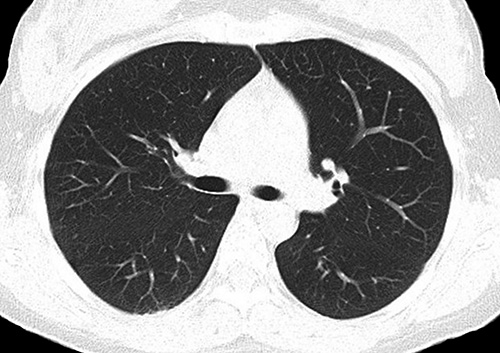 Computed tomography (CT) is a diagnostic procedure that uses specialized X-ray equipment to create cross-sectional pictures of the body. The CT computer displays these pictures as detailed images of organs, bones, and other tissues. This procedure is also called CT scanning, computerized tomography, or computerized axial tomography (CAT).
We have seven CT scanners in operation at University of Utah Health, including two cutting-edge clinical and research scanners, the Siemens Somatom Force and the Siemens Multitom Rax (see below).
Siemens Somatom Force
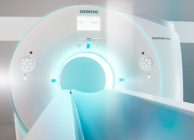 In December 2017, University of Utah Hospital installed the next generation of CT technology that allows for lower doses of radiation exposure and even quicker exam times. The Siemens Somatom Force is a dual-source CT scanner, meaning it has two radiation tubes and detectors that rotate around the patient. They are completely new X-ray tubes with high current, low voltage, a larger detector, and faster speed of rotation. What's more, the system uses great advances to the software that interprets the data.
These improvements mean that patients who have a concern for radiation dose or are unable to hold their breath for a long time - like children or those with lung and heart conditions - can now receive important diagnostic CT scans without as much risk and with good image quality from a much shorter scan time.
Siemens Multitom Rax
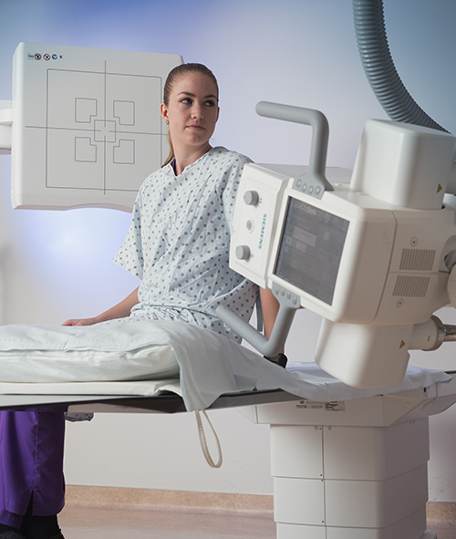 In 2016, University of Utah Hospital installed the world's first Twin Robotic X-ray system, which can perform exams from emergency medicine to pain management and orthopedics, and from conventional 2D radiography to fluoroscopy exams and angiography applications.
The unique open design of the Multitom Rax Twin Robotic X-ray system features a height-adjustable patient table and two independent, ceiling-mounted robotic arms for the X-ray tube head and the flat-panel detector. This allows almost unlimited positioning freedom anywhere in the room. Both robotic arms can be moved into position automatically or manually with servo motor support to make fine adjustments. While one robotic arm moves the X-ray tube, the other arm carries the 17” x 17” flat panel detector, which can acquire static, dynamic, and Real 3D sequences.
The Multitom Rax Twin Robotic X-ray system enables, for the first time, the acquisition of 3D images under the patient’s natural weight-bearing condition – whether the patient is seated, lying down, or standing. Images acquired in the natural standing position are essential because the knees, pelvis and spinal column appear differently when the patient’s body weight is applied compared to when the patient is lying down.
Micro CT
MicroCT scanners are used in biological and other basic research to investigate the anatomy of, among other things, experimental animals. Using non-invasive imaging techniques to analyse the response of experimental animals has enormous advantages to the investigator and, most of all, to the animal. For example, measurements of tissue development can be made without sacrificing the subject and without pain. Repeated measurements may thus be made over time in the same animal, providing more accurate information and requiring fewer animals.