
PET

PET
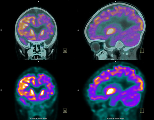 Positron-Emission Tomography (PET) is a nuclear medicine technique using a camera that captures images of how the human body functions. Compounds normally existing in the body, like simple sugars, are labeled with radioactive tracers, and are injected into the body intravenously. The tracer emits signals, which are recorded by the scanner as the tracer travels through the body and/or collects in targeted organs. A computer reassembles the signals into actual images, which then show biological maps of normal organ function and failure of organ systems in disease.
The basic steps of PET imaging include radioisotope production, radiopharmaceutical chemistry, tomographic acquisition, and image re-construction.

PET Scanners at the U
The University of Utah has five PET or PET/CT scanners in operation: