
SPECT

SPECT
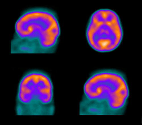
Single photon emission computed tomography (SPECT) is a nuclear medicine imaging technique using gamma rays. SPECT provides a 3D image of how a biological process functions in a patient. A gamma-emitting radiotracer is first injected into the bloodstream of the patient. Radiotracers are made to both emit a radioactive signal, which can be detected on the scanner, and to bind to specific molecules in the body. Patients wait a specific amount of time for the radiotracer to travel to the part of the body that physicians want to image, and then patients go to the scanner, where a gamma-camera captures how their body uses the radiotracer.
 SPECT images are different from the X-ray CT images. The X-ray CT uses external X-rays to image patient's anatomical structure, while SPECT uses injected internal gamma rays which emit a small amount of radioactive signal from the inside of a patient. The radiotracer is usually removed harmlessly from the body within 24-36 hours.
SPECT is similar to PET (Positron Emission Tomography) in its use of radioactive tracer material and detection of gamma rays. In contrast with PET, however, the tracer used in SPECT emits gamma radiation that is measured directly, whereas PET tracer emits positrons which annihilate with electrons up to a few millimeters away, causing two gamma photons to be emitted in opposite directions. A PET scanner detects these emissions "coincident" in time, which provides more radiation event localization information and thus higher resolution images than SPECT (which has about 1 cm resolution). SPECT scans, however, are significantly less expensive than PET scans, in part because they are able to use longer-lived more easily-obtained radioisotopes than PET.
During SPECT data acquisition, a gamma camera is used to acquire multiple 2D images (also called projections), from multiple angles. A computer then uses a sophisticated algorithm to "reconstruct" a 3D image. This dataset may then be manipulated to show thin slices along any chosen axis of the body, similar to those obtained from other tomographic techniques, such as MRI, CT , and PET .
To acquire SPECT images, the gamma camera is rotated around the patient. Projections are acquired at defined points during the rotation, typically every 3-6 degrees. In most cases, a full 360 degree rotation is used to obtain the best reconstruction. The time taken for each projection is also variable, but 15-20 seconds is typical. This gives a total scan time of 15-20 minutes.
Multi-headed gamma cameras can provide a faster scan. For example, a dual headed camera can be used with heads spaced 180 degrees apart, allowing 2 projections to be acquired simultaneously, with each head requiring 180 degrees of rotation. Triple-head cameras with 120 degree spacing are also used.
Cardiac gated acquisitions are possible with SPECT, just as with planar imaging techniques such as MUGA. Triggered by Electrocardiogram (EKG) to obtain differential information about the heart in various parts of its cycle, gated myocardial SPECT can be used to obtain quantitative information about myocardial perfusion, thickness, and contractility of the myocardium during various parts of the cardiac cycle; and also to allow calculation of left ventricular ejection fraction, stroke volume, and cardiac output.
Because SPECT permits accurate localization in 3D space, it can be used to provide information about localized function in internal organs, such as functional cardiac or brain imaging.