 |
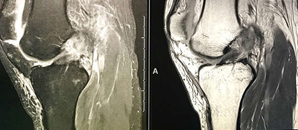
| Two MRI views of a torn ACL in the knee. Photo courtesy University of Utah Department of Radiology and Imaging Sciences. |
By Michael Mozdy
Sports fans cringe when we see one of our players go down on the court or the field. Seeing a basketball player hobble to the bench, clutching at his ankle, a skier sledded off with a stabilized knee after a bad tumble, or a football player taken out in an ambulance with his head and neck held perfectly still makes us empathize with what might be career-limiting injuries for high-profile athletes who push themselves to the physical breaking point.
What each of these players has in common is that they rely on a team of skilled physicians at University of Utah Health to take care of them when injuries strike. The University of Utah Orthopaedic Center is the official sports medicine provider for the Utah Jazz, U.S. Olympic and Paralympic athletes training and competing in Utah, the Salt Lake Bees, the Salt Lake City Stars (NBA G-league), and of course the University of Utah Utes athletic teams.
During and after sporting events, our orthopedic physicians work with team trainers to assess injuries, and when something serious or mysterious is suspected, the job falls to our radiologists to interpret studies and help determine the outlook for the athlete.
The Joint’s the Thing
Athletes push and punish their bodies, and this stress greatly increases the chances of sprains, bruises, tears, and breaks. Joints are where bone, muscle, cartilage, ligaments, and tendons attach to one another and also provide the magical ability for us to pivot and bend in so many ways. Because they are focal points for our bodies’ complex anatomy, joints are common sites of injury: ankle, knee, shoulder, wrist, etc. With so much happening in a joint, however, it can be tricky determining exactly what anatomy is damaged and how to best treat it.
We have a phalanx of skilled orthopedic physicians attending to these athletes – Travis Maak, MD, David Petron, MD, Stuart Willick, MD, and Patrick Greis, MD, just to name a few. In addition to a physical exam, these docs will often partner with our musculoskeletal radiologists to diagnose, treat, and monitor these injuries.
 |
| Chris Hanrahan, MD, Section Chief for Musculoskeletal Radiology. |
Chris Hanrahan, MD, is the Section Chief for Musculoskeletal Radiology, and leads a dedicated team of experts who read scans day in and day out at the Orthopaedic Center. After completing their medical degrees, each physician has 4 years of radiology training and an additional year of subspecialty training in musculoskeletal imaging, particularly orthopedic MRI. Add to this their many years of experience at the Orthopaedic Center, and it’s no wonder so many world-class athletic programs trust us.
All of this expertise is needed when athletes experience pain and limited movement because MRI is the best tool to reveal the extent and location of soft tissue injury. When money and careers are at stake, the big question is if athletes should stop playing and practicing in order to recover from an injury. The guidelines for “return to play” in the case of concussions have been well defined in recent years, but for joint injuries, the criteria are less clear and more flexible. It often comes down to a discussion between player, trainer, physician, and coach. Understanding the extent of the injury is crucial for this discussion, and a radiologist’s MRI report plays a major role.
Sleuthing Stories in Scans
 |
| Megan Mills, MD, faculty in the Department of Radiology and Imaging Sciences. |
“Everyone wants x-ray vision, but I’d rather have MRI vision,” claims Megan Mills, MD, one of our musculoskeletal radiologists. She’s not exactly joking. She explains that while x-ray images may show fractures and massive ligament tears can be clinically obvious, these aren’t the only injuries that could be causing pain. “There could be subtle fractures, cartilage injuries, meniscal or labral injuries,” Mills explains, not to mention what she calls “loose joint bodies,” or little calcifications that just hang out in your joint, “and only MRI is going to show you that.”
These “secondary findings” can greatly impact the course of treatment and recovery. Take a common (and season-ending) injury: the ACL tear. The anterior cruciate ligament (ACL) is a major ligament in your knee connecting your thighbone to your shinbone, and over 200,000 people tear their ACL each year in the USA. The musculoskeletal radiologists assert that it’s vital to examine the smaller stabilizers (ligaments and tendons) around the ACL to see if they’re injured as well. If these injuries aren’t found and fixed, the knee won’t be stable in a side-to-side motion, and an athlete might find herself back on the operating table a second time to fix this. A detailed MRI interpretation guides surgery and therapy to properly heal an athlete.
MRI is a sensitive tool for sports medicine because it can show clear distinctions between all of the anatomy – skin, bone, muscle, tendons, etc. Even some bone fractures, which you might think x-ray is sufficient to show, are better found with MRI. Metatarsal stress injury in the foot – often subtle on x-ray – can be found on MRI immediately. This is an important one because runners and football players could either let a partial stress injury in this bone heal or unknowingly create a complete (and much more severe) fracture through the bone by continuing to play on it if it is not identified. MRI can also find acute edema (fluid and swelling in tissue) within hours, and muscle changes nearly immediately.
 |
| Sarah Stilwill, MD, faculty in the Department of Radiology and Imaging Sciences. |
Our radiologists go beyond simply catching these findings and look for injury patterns in their work. “It’s a story,” declares Sarah Stilwill, MD, another musculoskeletal radiologist in the department. “We do some sleuthing and trace the apparent injury back to the action of play (which is the fun part!) to determine how it happened. Knowing the patient’s mechanism of injury ensures you’re not missing what else might be impacted.” Stilwill explains that the patient history of the injury is the most important piece of information when they’re interpreting an athlete’s scan.
The radiologists have the highest praise for their MRI technologist colleagues, who not only collect patient histories when they come in for the scan, but ensure patient safety, appropriate positioning, and expert imagine technique when performing scans. “If you have the story,” Stilwill continues, “you’re focusing on the site of pain, but also other structures that would go along with that injury pattern.” To fully probe the potential implications of these injuries, the radiologists have to be experts in anatomy and understanding how different musculoskeletal structures work together for joint stability.
Direct Treatments from Your Radiologist
While MRI is a favorite tool, the radiologists also examine x-ray and CT scans on a daily basis. And in addition to diagnosing injury, they use their scanning equipment to guide procedures and treatments.
Rather than blindly sticking a needle into a joint and fishing for the correct point of treatment, our radiologists use fluoroscopy (x-ray) or ultrasound in real-time to see the structures and deliver treatments precisely where they need to go. Because the core of their work is noticing minute differences in soft tissue and how vasculature, cartilage, muscle, and bone relate, they have a great sensitivity for being able to accurately apply treatments.
Two common procedures are biopsies and steroid injections. Biopsies are used to determine if a lesion is cancerous, and steroid injections provide pain relief to enable physical therapy and healing. Hanrahan describes an instance where a steroid injection helped diagnose a hard-to-find pain point in a basketball player’s ankle. “After having ankle surgery, he had persistent pain and we couldn’t tell why,” Hanrahan explains. A steroid injection adjacent to a tiny bone spur provided immediate relief, and helped to pinpoint the problem “After a second surgery on the bone spur, he returned to play and was able to have a good career.”
Both biopsies and injections require a long needle, navigation around complex anatomy, and a steady hand. The steady hand comes from intimate knowledge of anatomy and plain old practice. As the only training ground for physicians between Denver and the West coast, the U of U needs to show resident physicians how to capably accomplish these procedures. To that end, Mills has applied for a grant to print realistic 3D models of joints – made with materials that realistically simulate skin, muscle, tendons and bone – in order to train residents without practice on a live patient. Innovative techniques like these make patients safer and improve the quality of the care we provide.
 |
| The Musculoskeletal Radiologist team (left-to-right): Sarah Stilwill, MD, Anna McGow, MD, Chris Hanrahan, MD, Megan Mills, MD. |
Pushing the Understanding of Sports Injuries
Despite our ability to visualize injuries, determining their impact on player readiness and the need for rest or treatments can remain elusive. The bone bruise is one such injury, and also one where we are attempting to expand our understanding by conducting research.
Bone bruises – what radiologists call bone marrow contusions – don’t show up on x-ray, but can be seen on MRI. Essentially, they happen when there is compression or a direct blow to the bone. It’s an injury that is less severe than a fracture, but it can cause pain, stiffness, and the inability to use joints effectively. More confounding for physicians, bone bruises can last up to 6 months. When athletes continue to pound their bodies, it’s difficult to know if a bone contusion on MRI is from months ago or is due to last night’s collision on the court. This also makes it difficult to recommend how long or whether an athlete should take a rest to allow for healing.
Maak and Hanrahan have applied for a grant from the NBA to study bone marrow contusions over the course of a year to better understand how they relate to overall performance, injury, and readiness to play.
“This type of study is badly needed,” says Hanrahan, “because the implications of multiple bone contusions on readiness to play is not clear.”
Hanrahan is deeply dedicated to providing the most state-of-the-art care possible. “We are constantly working to make sure we have top-notch equipment and fine-tuning our scans and procedures.” For instance, Hanrahan has partnered with Eugene Kholmovski, PhD, in the department’s Utah Center for Advanced Imaging Research to optimize MRI pulse sequences on nerve imaging studies. They now have images that are stunningly improved over the ones that are made with the MRI manufacturer’s settings for that scan.
All of this expertise and research makes the University Orthopaedic Center a magnet for programs that need serious care for their athletes. But isn’t it great to know that these awesome radiologists are the same ones who read the scans for you and I? From grandmothers to toddlers, it’s reassuring to know that this expertise is available to all.
Based on her years of observing the patterns of injury, Stilwill provides us with a final word: “Overall, people need to be more careful. The body recovers well, but it’s extremely fragile. It doesn’t take much to put your bones and muscles in peril. You constantly have to make sure you’re staying fit and active to keep how the body works strong.”
Truly words that matter to athletes and couch potatoes alike.