
The Force is With Us: Reviewing the Power of our Latest CT Scanner

Apr 17, 2018
By Michael Mozdy
A typical day in the life of a CT scanner at the hospital is a busy one: 20 chest scans, 35 head, spine and neck scans, 2-3 cardiac scans, 20 abdomen scans, 10 angiograms and 2-3 interventional radiology procedures. Many of these are ordered from the emergency department, but patients come from all across the hospital, both inpatient and outpatient areas.
 |
| Saeed Matinkhah, CT Manager |
“CT is the workhorse for emergency medicine,” acknowledges Saeed Matinkhah, CT Manager for the health system.
CT machines are so busy because the scans are quick, they provide physicians with useful diagnostic information, and they cost less than MRI or nuclear medicine studies. There is a tradeoff, however: like X-ray scans, CT machines project radiation through the body, which has some risks. For this reason, CT researchers and manufacturers have been working hard to achieve “ultra low-dose” scans.
In December 2017, the Radiology Department installed the newest technology – what they call third generation technology – from the manufacturer Siemens and brought ultra low-dose, extremely quick CT scans to University of Utah Hospital. This remarkable new machine came with a catchy name, too: the Somatom Force.
What Makes a Good Scan?
The best scans for medicine are ones that are:
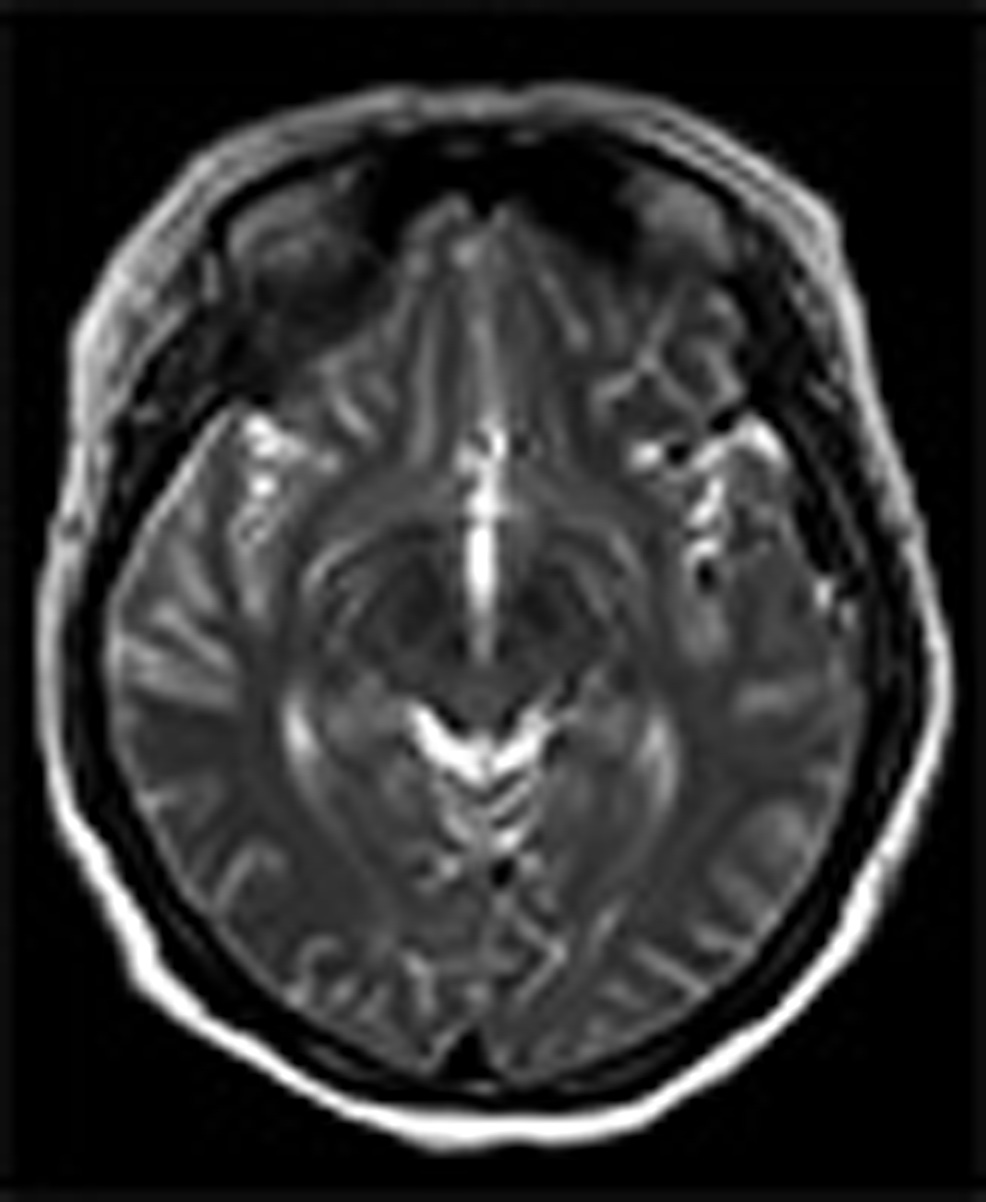
Detailed – thanks to the advent of the digital age, most of us are familiar with the difference between low resolution and high resolution pictures. In digital photography, low resolution means that the image becomes blurry or pixelated when you try to enlarge it, and you can’t see as much detail as a result. In imaging sciences, this is called spatial resolution, and it describes the size of a scan’s voxels (three-dimensional pixels). If a scan has high spatial resolution, then radiologists may see small details in the image.
|
|
| Photoshop manipulation of stock brain CT image – the image on the left has low spatial resolution (less pixels per inch) while the one on the right has high spatial resolution. | |
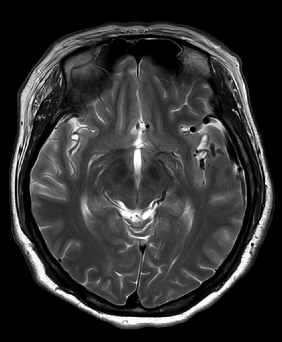
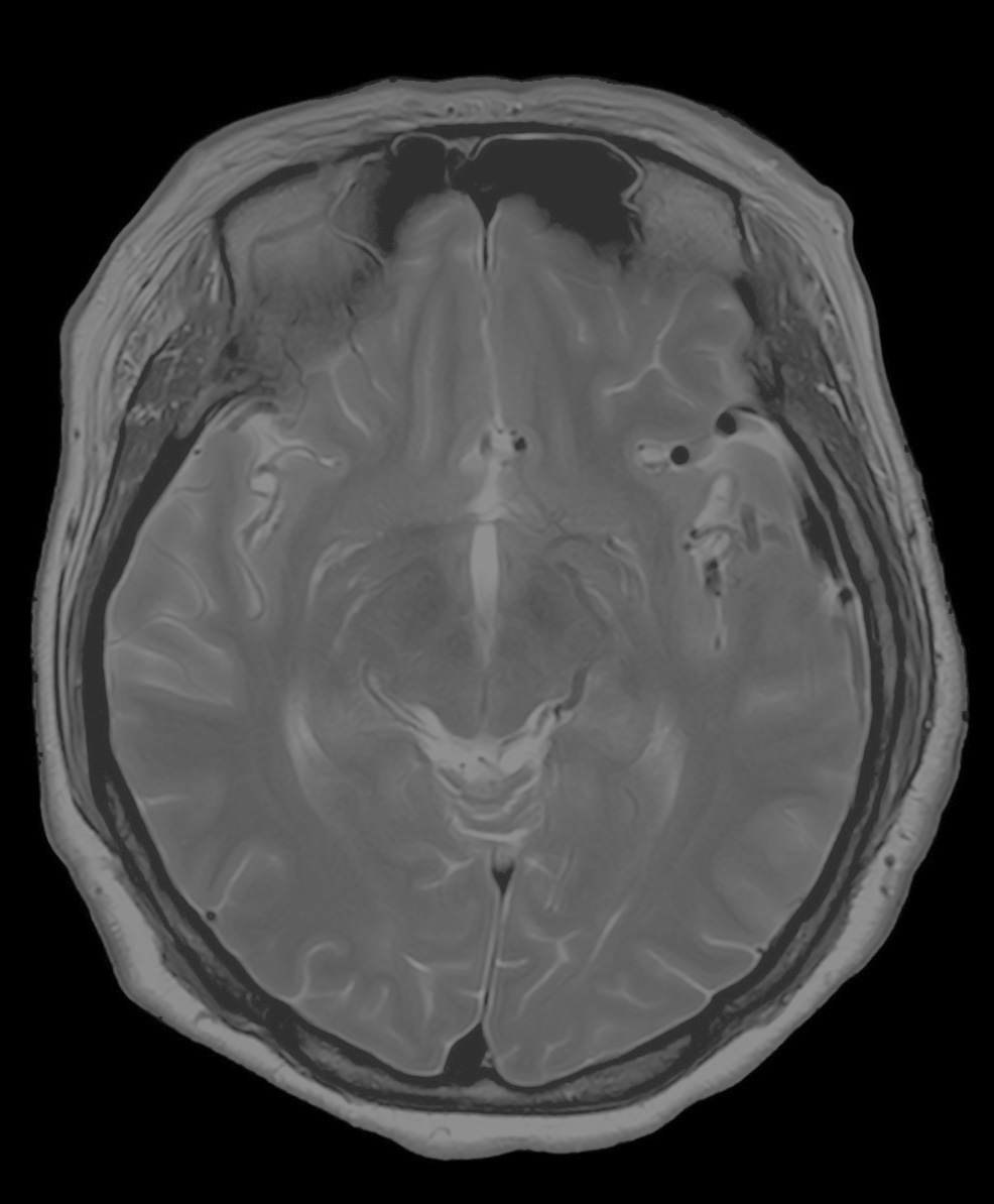
Clear – since CT, MRI, ultrasound, and nuclear medicine scans look at the inside of your body without actually cutting you open, the data is not the visible color spectrum. In the case of CT, the image displays how x-rays are absorbed and scattered by the different tissue types in the body, and this is a grayscale image. Even with good spatial resolution, if there is not enough contrast in the signal that is measured by the CT, the images could appear as a field of gray, white and black blobs. Good contrast resolution results in images that have clearly defined anatomy. Clinicians can increase contrast resolution by using a special dye called a contrast agent or through better detectors in the machine and software that interprets the data being collected.
|
|
| Photoshop manipulation of stock brain CT image – while the two images have the same spatial resolution, the image on the left has low contrast resolution while the one on the right has high contrast resolution. | |
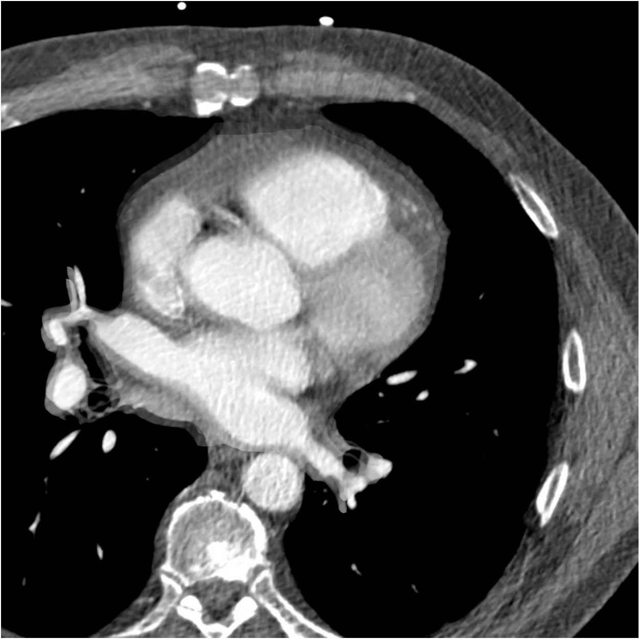
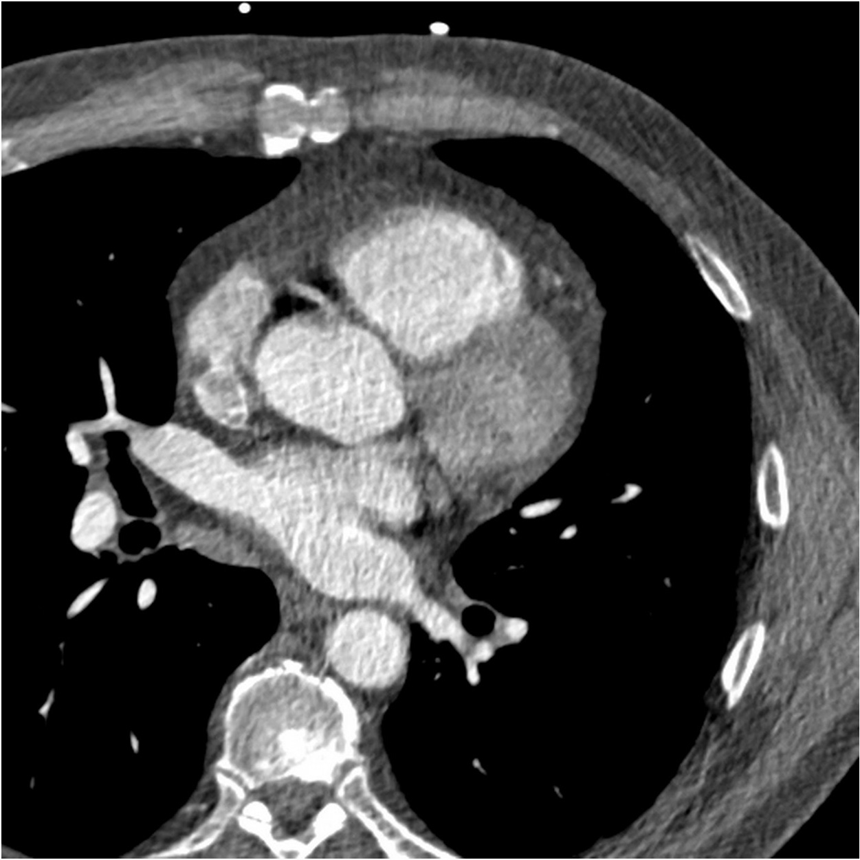
Quick – Even with high spatial and contrast resolution, images will become blurry if the scan can’t capture information quick enough. For example, it is difficult to scan a beating heart moving around in your chest or your lungs expanding when you breathe. This ability to capture things in a short time window, akin to shutter speed, is called temporal resolution. Other advantages of a quick scan are that patients prefer them and they’re less costly in terms of personnel time.
|
|
| Photoshop manipulation of stock cardiac CT image (cross section of the heart, showing separate chambers) – while the two images have the same spatial and contrast resolution, the image on the left has low temporal resolution, so the beating heart causes blurred “motion artifacts” while the scan is performed, while the one on the right has high temporal resolution and therefore better delineation of the structures. | |
The Power of the Force
CT scanners operate by having the radiation source and a detector rotate around the patient in a big donut-shaped ring called a gantry. The gantry is, in fact, packed with sophisticated equipment that is traveling around the patient at incredible speeds. Our new Somatom Force scanner is a dual-source scanner, meaning that it has two photon emitters across from two detectors – enabling twice the coverage in the same amount of time as a single source CT scanner.
 |
| Section Chief for Cardiovascular Imaging, Joyce Schroeder, MD |
Advances in the type of emitters, detectors, software, and the speed of the gantry mean that the Somatom Force excels in all three of the types of resolution imaging scientists like to optimize. Perhaps most importantly to patients, what this means is good contrast resolution with nearly half the amount of radiation during a typical CT scan. Most CT scanners already have very good spatial resolution – voxels about 0.5 mm3 – and this scanner improves upon this to achieve 0.3 mm3 voxels. Finally, the gantry can revolve around the patient in an unbelievable 0.25 seconds. This means that the entire heart, for instance, can be scanned in literally less time than a heartbeat. Patients who would have trouble remaining still (trauma patients, children) and those who would be unable to hold their breath (elderly, people with lung problems) can now receive an accurate CT scan without motion blurring in the images.
Our Section Chief for Cardiovascular Imaging, Joyce Schroeder, MD, has great things to say about the Somatom Force. “We can take care of our patients in a better way thanks to improved spatial and temporal resolution in a package that provides ultra low-dose and fast scans,” she praises. “We use less radiation and less intravenous contrast. It’s really a win-win scenario.”
Our Administrative Director of Clinical Services, Lisa Bakhsheshy, is happy to have the Somatom Force as one of the seven CT scanners in the system. “Any time you make an investment in major medical equipment, you want to ensure that it brings a clear benefit for your patients and your clinicians,” she explains. “and this scanner provides clear benefits for everyone.”