By Michael Mozdy
Parents expecting a child encounter a vastly different world today than just 20 years ago. Perhaps the most striking example of our commercialized, child-focused culture is the service of taking portrait pictures of your unborn fetus. It sounds a little premature, to say the least, but thanks to the incredible advances in ultrasound reconstruction algorithms and resolution, it is an irresistible purchase for parents anxious to see their little one in their arms.
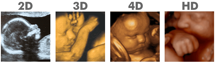
The newest ultrasound images advertised by a commercial fetal ultrasound company.
Yet it’s more than just a pre-natal photo booth novelty. “We interpret 3D and 4D ultrasound studies every day in our practice,” states Paula Woodward, MD, Radiology and Imaging Sciences faculty member, holder of the David G . and Marcia R. Presidential Endowed Chair in Radiology, and co-author of the best-selling textbook Diagnostic Imaging: Obstetrics, 3rd Edition. “It’s an indispensable tool for diagnosing and characterizing potential defects and deformities.”
Woodward and her fellow faculty ultrasound expert (and textbook co-author), Anne Kennedy, MB, BCh, BAO, have been advancing ultrasound imaging for over 30 years, and they’ve never been as excited about its diagnostic utility as this moment in time.
Flexible, Real-Time Problem Solving
Ultrasound is a workhorse for many medical specialties, notably maternal and fetal medicine and cardiology (yes, that echocardiogram is an ultrasound machine). The low-energy sound waves it delivers to body tissues are very safe – in fact, generally considered completely harmless. The hand-held ultrasound device – called a transducer – glides over the patient’s skin and is re-positioned easily by the operator thanks to a gel put on the patient’s skin. The gel eliminates any air pockets that could disrupt the images because they don’t conduct sound that well. Ultrasound machines are affordable (as low as $25,000, compared to $2 million for an MRI machine), and the exams provide very close to real-time imaging, providing immediate information to physicians and patients alike.
All of these advantages have made ultrasound the most used imaging around the world – by a long shot. Developing countries can’t afford the cost needed to purchase and maintain more complex imaging like PET, CT, and MRI. First-world countries in Europe have also embraced ultrasound, not for a lack of resources, but because it keeps the cost of health care down and provides quick diagnoses of so many disease states.
Even here in the technology-obsessed United States, ultrasound has been gaining footing in primary care doctor’s offices around the country. It’s much better customer service to check out your patient’s knee with the portable ultrasound machine in your office to rule out one thing or another rather than send that patient through scheduling a separate visit for the ultrasound at another location and waiting for those results to be interpreted by a radiology team.
“It’s targeted problem solving,” explains Kennedy.
Yet while family practice docs, OB/GYN physicians, cardiologists, and more are finding that ultrasound is so approachable, affordable, and helpful to perform and interpret themselves, Kennedy and Woodward still see a very prominent role for radiologists. “Our training as radiologists gives us an eye for reading studies beyond a suspected diagnosis,” Kennedy says. “Those who only use ultrasound now and then or for a limited area of interest might, for instance, look thoroughly at the gallbladder but completely miss the liver tumors sitting there on the exam.”
Kennedy and Woodward spend most of their work hours in radiology reading rooms, but have the opportunity to work directly with patients when a study done by a sonographer is inconclusive. That’s when they see the real value of ultrasound shine.
“It’s targeted problem solving,” explains Kennedy. “You have the opportunity to try different transducers, different scan planes, and narrow down from a giant list of diagnoses to a specific diagnosis. Ultrasound has the potential to be like that in every case.”
Woodward and Kennedy appreciate the time spent with patients doing real-time problem solving because it provides more than technical benefits. “It’s one of the few areas in radiology where you are actually there talking to the patient,” explains Woodward. “We often get a better patient history than the referring physician.” Some of the clues dropped by patients guide them to a better exam, for instance, knowing that some trauma happened or that their pain had been recurring over time. Like medical detectives, they interrogate the scenario with their tools, their words, and their brains, fitting together the evidence as completely as possible to arrive at a more specific diagnosis for the patient.
It’s similar to the way ultrasound is used by radiologists in our Breast Imaging section. When women are called back for additional imaging because something suspicious showed up on their breast screening exam or they have a lump, ultrasound is an invaluable tool used to examine an area of interest further. It can quickly and easily help the radiologist to determine if an abnormality warrants a biopsy or if it’s benign.
 |
| Nicole Winkler, MD, assistant professor in the Department of Radiology and Imaging Sciences and Breast Imaging Fellowship Director. |
Nicole Winkler, MD, faculty member and Breast Imaging Fellowship Director agrees that ultrasound is a flexible, safe, and extremely helpful tool in her work. “We use ultrasound guidance for many breast-related procedures,” she says. “It is great for biopsies because we are able to watch in real-time as our needle traverses a mass or abnormal area to ensure we biopsy the intended target.” They also use ultrasound for treatment of breast infections to guide the drainage of breast abscesses and seromas.
Another tricky procedure is surgically removing breast cancer or other abnormalities when they cannot be felt externally by surgeons. Winkler explains that radiologists use ultrasound to locate the cancer prior to surgery and place a tiny radioactive seed within the tumor that can be detected intraoperatively by the surgeon using an external probe with ±1mm accuracy. This enables a much more accurate and successful procedure.
A further aspect of ultrasound is that it can provide information about tissue-stiffness properties in the breast – called elastography. This can be used by radiologists differentiate types of tissue they see, such as a cyst containing debris from a solid mass.
Winkler, like Kennedy and Woodward, can’t imagine a successful modern radiology practice without the extensive use of ultrasound.
Spreading the Gospel of Ultrasound
Woodward and Kennedy are on a mission to make sure the radiology residents they train learn these valuable problem solving skills. Having worked together for 25 years, they make a great team, and are self-avowed “twins,” finishing each other’s sentences frequently.
Woodward went to medical school with help from the Air Force, where she did her residency. Before taking her radiology board exams, she was appointed the chief of ultrasound at a 1000-bed hospital. “I didn’t choose ultrasound, I was ordered to it,” she laughs. She quickly learned to love the subspecialty. After this experience, she came to the University of Utah, and her first fellow in the OB radiology section was none other than Anne Kennedy.
Kennedy was initially an internal medicine physician in England, but began spending time with radiologists, who piqued her interest in the field. “I was particularly interested in ultrasound because of the unique way the study can be customized to answer a specific clinical question,” she relates. Kennedy and Woodward soon found that they fed each other’s interest in and love of diagnostic ultrasound.
Their professional partnership has notably focused on educating others on the power of their tool. The 3rd edition of their textbook, Diagnostic Imaging: Obstetrics, came out last year. It’s the medical book equivalent of a New York Times best seller, with a perfect 5-star Doody review (only 8% of the thousands of textbooks annually reviewed by Doody’s 5,000+ academically-affiliated clinicians receive this score). They are now working on a book for sonographers, a technician-level book focused on their needs.
Knowing that the technique and subtlety of their subspecialty is best imparted in person, they both lecture extensively nationally and internationally. They are well-known at the world’s premier radiology association – the Radiologic Society of North America (RSNA). For the past two years they’ve presented the “OB Case of the Day” at the RSNA meeting, which is five days’ worth of interactive case studies taken by many thousands of clinical radiologists. What’s more, this year their educational exhibit “Doppler for Babies,” won a magna cum laude award (only 2.6% of exhibits achieved this level of recognition).
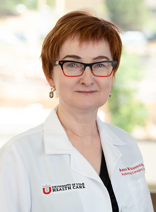 |
| Anne Kennedy, MB, BCh, BAO, Professor of Radiology and Imaging Sciences. |
Locally, Kennedy is a fetal echocardiography conference founding organizer. This conference is offered affordably so people from community ultrasound clinics can attend and learn how to refer patients when necessary.
Perhaps the most impactful and deep training they provide, however, is given to radiology residents at the University of Utah. They drill into their residents three concepts: anatomy, scan technique, and differential diagnosis. “It’s extremely important to us,” says Kennedy, “that they learn how to perform ultrasound studies and talk with patients.” The objective is a very focused report that can rule out lots of things, narrow down the possible diagnoses, and provide a recommendation of what to do next. “There’s nothing worse than a radiology report that lists a million things as a possible diagnosis,” proclaims Kennedy. “We tell residents to read over their reports and ask if they helped guide the management of the patient.”
Unsolicited, departing senior neuroradiology fellow Atul Mallik, MD, PhD, testifies to the impact such careful training can have. After four residency years and two more as a neuroradiology fellow, Mallik is busy finishing his research and clinical duties before going to his first faculty position at Loyola University in Chicago. Amid the heavy workload in his last month, he noted a bright spot in his week: “I got to perform an ultrasound study today,” he said with a smile, “It was really nice to get to talk with the patient and troubleshoot what was going on in a hands-on way.”
Mallik is not alone in his feelings – Kennedy and Woodward often hear back from alumni of the residency program that they have come to appreciate knowing how to perform these studies. Kennedy and Woodward are upholding a more traditional, almost artisanal approach to diagnosing disease, and the positive affect of their educational work will thankfully be felt for generations to come.
But just because it’s artisanal doesn’t mean it’s not cutting edge.
Surprisingly Sophisticated Technology
“If you look at images from the 1990s compared to now, the quality is astoundingly better,” Kennedy says enthusiastically. And while we might be used to thinking of CT and MRI changing drastically in the past 20 years, she believes that ultrasound has improved even more than those modalities.
 |
| Paula Woodward, MD, Professor and David G . and Marcia R. Presidential Endowed Chair in Radiology. |
Because the resolution and software algorithms have attained a new level of accuracy, much more power resides in the hands of the operator. “It is the ultimate multiplanar modality,” Woodward declares. “You can do obliques literally at the turn of your hand.” What Woodward means is that it’s not difficult to change the plane of view in a multitude of ways to visualize the anatomy differently. These “oblique views” can provide clinicians with valuable data that something like an x-ray never could, and something like an MRI or PET study would take hours and hours of time to compile and go through. This underscores the need to sufficiently train both sonographers and radiologists how to harness the power of the ultrasound exam for visualization and diagnosis.
And that’s not the only news about ultrasound. European countries are regularly using microbubbles filled with gaseous sulfur hexafluoride for contrast-enhanced ultrasound. As we’ve learned, gases interfere with ultrasound signals, and the microbubbles show up clearly under ultrasound. They help visualize blood flow and some disease states just as well as contrast-enhanced CT and MRI. Excitingly, patients with kidney problems who can’t receive CT or MRI with contrast because that type of contrast is processed through the kidney can, in fact, undergo contrast-enhanced ultrasound. The microbubbles are actually passed out of the body through the lungs.
This technology isn’t available for patients in the United States because the FDA has been holding up use of microbubbles here. However, Kennedy and Woodward’s colleague Tom Winter, MD, Chief of Abdominal Imaging, is evaluating renal transplant patients with the help of contrast-enhanced ultrasound as part of a funded research project.
Contrast-enhanced ultrasound is invaluable in cases like portal vein thrombosis, which is a major concern with liver disease. It can be difficult to diagnose with conventional ultrasound, so these sick patients must be transported through the hospital to receive a CT. With contrast-enhanced ultrasound, however, it’s a simple diagnosis, safer, less expensive, and can be done at the patient’s bedside.
Our ultrasound service has more than doubled in the last 10 years, mostly due to outpatient ultrasound studies done in our community clinics. The entire abdominal section contributes their vast experience and expertise in interpreting these often challenging cases. They were joined by a new Women’s Imaging fellow this past year, Scott Parker, MD, and will have another such fellow, Elaine Pigman, MD, in 2018-19.
We are very proud of our busy ultrasound imagers and the great expertise they bring to our region and beyond. We hope that they are an inspiration to many generations of radiologists to continue the work of medical detective, attentive to their patients’ stories and the sensitivity of their tools.