By Michael Mozdy
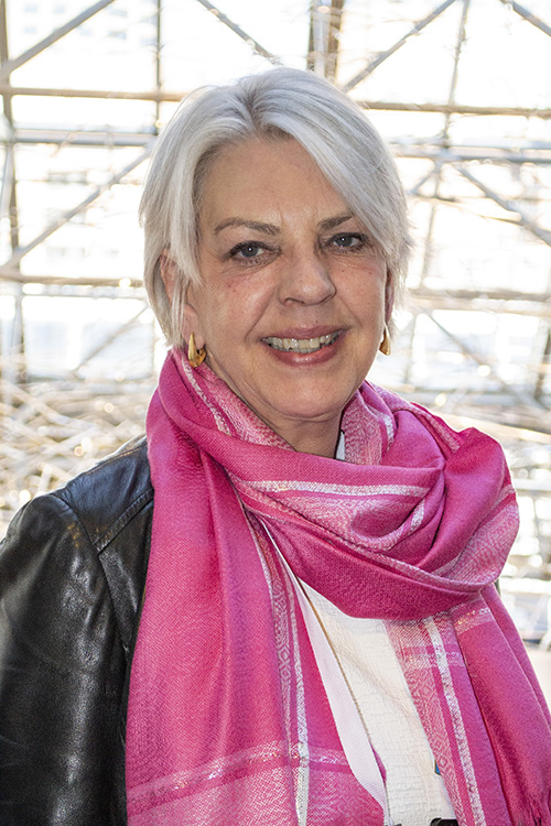
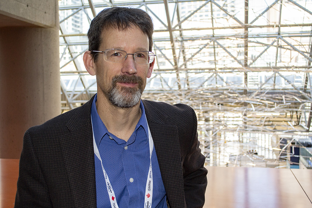
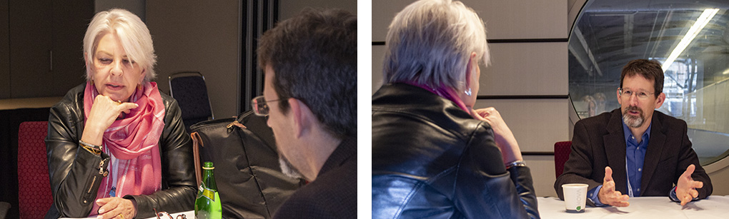
We recently had the opportunity to sit down with the President of the International Society of Magnetic Resonance in Medicine (ISMRM), Pia Sundgren, MD, PhD, during the ISMRM 2019 Annual Meeting in Montreal (read more about Sundgren at the end of this article). Ed DiBella, PhD, the Director of the Utah Center for Advanced Imaging Research (UCAIR) had an engaging conversation with her about the state of MR research and clinical practice.
What’s exciting to you right now in the field, especially something translatable?
Pia - In terms of the technical aspects of our field, I would say fast imaging techniques, which increase patient throughput through your scanner and also increase patient comfort since the scanning doesn’t take as long. Yet you’re still acquiring good images…
Ed – …or even better images because you’re moving faster and acquiring less so there can be less motion artifact.
Pia - The only caveat here is that while we are scanning more patients in less time, we need more radiologists on the other end to report on the study. Manpower becomes an issue, but this is where machine learning may come in. You can reduce the time you as a clinician need to spend looking for that extra MS plaque that has showed up. You can shorten the time you need to spend reviewing images.
Ed – I came to the meeting sort-of up in the air on what the impact of machine learning might be, but the plenary talks were amazingly convincing that the impact is very soon. Especially with segmentation and tools to help reading.
 Pia – I am definitely an advocate for using machine learning in the appropriate manner – we clinical radiologists are still the final say in terms of what’s a lesion and what’s an artifact. It’s just going to modify our way of working and help us in many ways – I’m not worried that we’ll be out of work! And there will be areas of medicine where machine learning will just not apply, where we could not have enough cases to train the algorithm. For instance, rare metabolic diseases in children where you might only see one case in your lifetime. Here you need an expert team of people.
The other important issue is that if you’re not very careful, you’ll have “garbage going in” to train a system, and you’ll get “garbage out.” So, radiologists and MR physicists need to be involved in what data is going into the system – what information do you need to ensure is present to result in a good model for segmentation or lesion detection? Take for example a data set of 1,000 MS patients. You could have 10 patients with sarcoidosis, which can look very much like MS, but if they go into the data set, 10-20 cases can disrupt the data set. I’m still a little bit of a skeptic in machine learning when it comes to not knowing who created the data set. Did you validate the data going in? That is something that can be easily overlooked.
Ed – Another concern is that there are so many different platforms and MR sequences, so there is a huge challenge in making sure the data going into your training set is equivalent to each other. Data standardization and data harmonization have become subfields of machine learning.
Pia – yes, and even from vendor to vendor – is a T1 scan on a Siemens scanner the same as one on a Philips scanner? We treat them as the same, yet the image contrast may vary. Some lesions will be more evident on one machine compared to another.
Ed – how do we try to push data harmonization?
Pia – we as individuals and our professional societies need to have these discussions with vendors and manufacturers.
Ed – This data harmonization problem reminds me of a criticism of MRI in general – that there are too many knobs and controls. You have to set 500 parameters! It’s a big difference between this and, say, “push-button CT” where you just push the button and the scan is done.
Pia – But if MR was push button, we run the risk of oversimplifying important concerns What pulse sequences and other parameters are being used when that button is pushed? Probably, most people don’t know or care, but that’s a problem. At our institution, we put a lot of effort in teaching the people who execute the scans what is happening when that “button” is pushed.
Ed – Absolutely. In my field of cardiac MRI, it’s clear to us that a good radiologic technologist equals good data.
Pia – Back to the question of what is interesting and new: on a clinical level, I am intrigued by techniques that avoid using gadolinium. Techniques like CEST, amide proton transfer imaging, and more advanced diffusion techniques that look at a cellular level, these help us better understand the inner structure of a lesion or a tumor. You can then correlate these findings with histopathology.
I work a lot on brain tumors, and always collaborate with three groups: MR physicists, neurosurgeons, and radiographers to see how we can help the clinicians pick the right area for surgery. We might not be able to do microscopic histopathology, but we can show that it’s not just a lesion, it’s not just a tumor –there are specific regions and we can help guide the neurosurgeon on interventions.
Ed – To tie back in with artificial intelligence, as you show how those patterns are more subtle, deep learning can help wade through this data.
Pia – Yes, and there are other factors MR is beginning to uncover on this fine scale that can guide treatment. The same grade of a tumor can behave completely differently based on molecular and genetic factors. If you can catch that at an early stage, maybe they can get the right treatment from the start instead of a second-line treatment.
Are there some technologies like 7T and PET/MRI that may not ever translate to broad clinical practice?
Pia – 7T is very expensive, I can’t see that it’s going to replace 3T around the world, although I feel like it has clinical value today in areas like epilepsy, lesion detection, trauma, injuries in the brain – varieties of 7T techniques may provide insight beyond basic morphology. PET/MR is great because of the simultaneous action, but the machine and the PET tracers are very expensive. It might not be sitting at every clinic in the future, and maybe it shouldn’t be. Maybe just at big hospitals or specialty centers where you can have a big enough patient cohort to justify its use and cost.
Ed – I’d add that it’s nice to see people working on 7T and PET/MRI because there is some technology trickle down. The problems are harder, and if you solve them, 7T solutions can benefit 3T systems.
Pia – You’re totally right. We should also know that 7T is not easy to implement, there are a lot of challenges. But when you get the system to work, you have the possibility to do more things there and then maybe bring it back later to the 3T environment.
Ed – Can I ask a different question? What’s been the best part of being the ISMRM president?
Pia – I think it’s the opportunity to be allowed to make an impact, to bring ideas for what we can do and how we can grow as a society. Last year, we had some great discussions about diversity, inclusion, and professional conduct. Members say it’s an open, safe, inclusive and diverse society; we’re going in a direction the members like. You meet a lot of people and friends from different parts of the world, and you feel appreciated for the work that ISMRM is doing.
Ed – The society is in a great place and this is a very exciting meeting. I want to add my compliments.
Pia – I want people be proud of being an ISMRM member and spread the word. The interaction between students, postdocs, trainees, MR physicists, clinicians, radiographers in the same setting, it’s pretty unique. Not many societies have this.
Professor Pia Maly Sundgren is the head of the Department of Radiology, Clinical Sciences/Lund, Lund University in Sweden, Co-Director for Lund University BioImaging Center, and senior consultant in neuroradiology at Skåne University hospital, LU, Lund. Her research focuses on advanced MR imaging and conducts imaging research focused on monitoring treatment response and effects of radiation in brain tumors, advanced imaging in autoimmune diseases with special focus on SLE as well as in pain conditions. Her research is conducted both at 3T and 7T. For advanced brain MRI she is an opinion leader and internationally recognized. Sundgren has published over 170 papers and reviews, over 210 scientific abstracts, three books and several book chapters and have given over 300 international invited lectures and workshops worldwide.