By Michael Mozdy
|
Printable Instructions for Health Care Providers For in-house printers/copiers (11x17") For print shops (with full bleeds, for trimming & more professional look - we recommend also laminating)
|
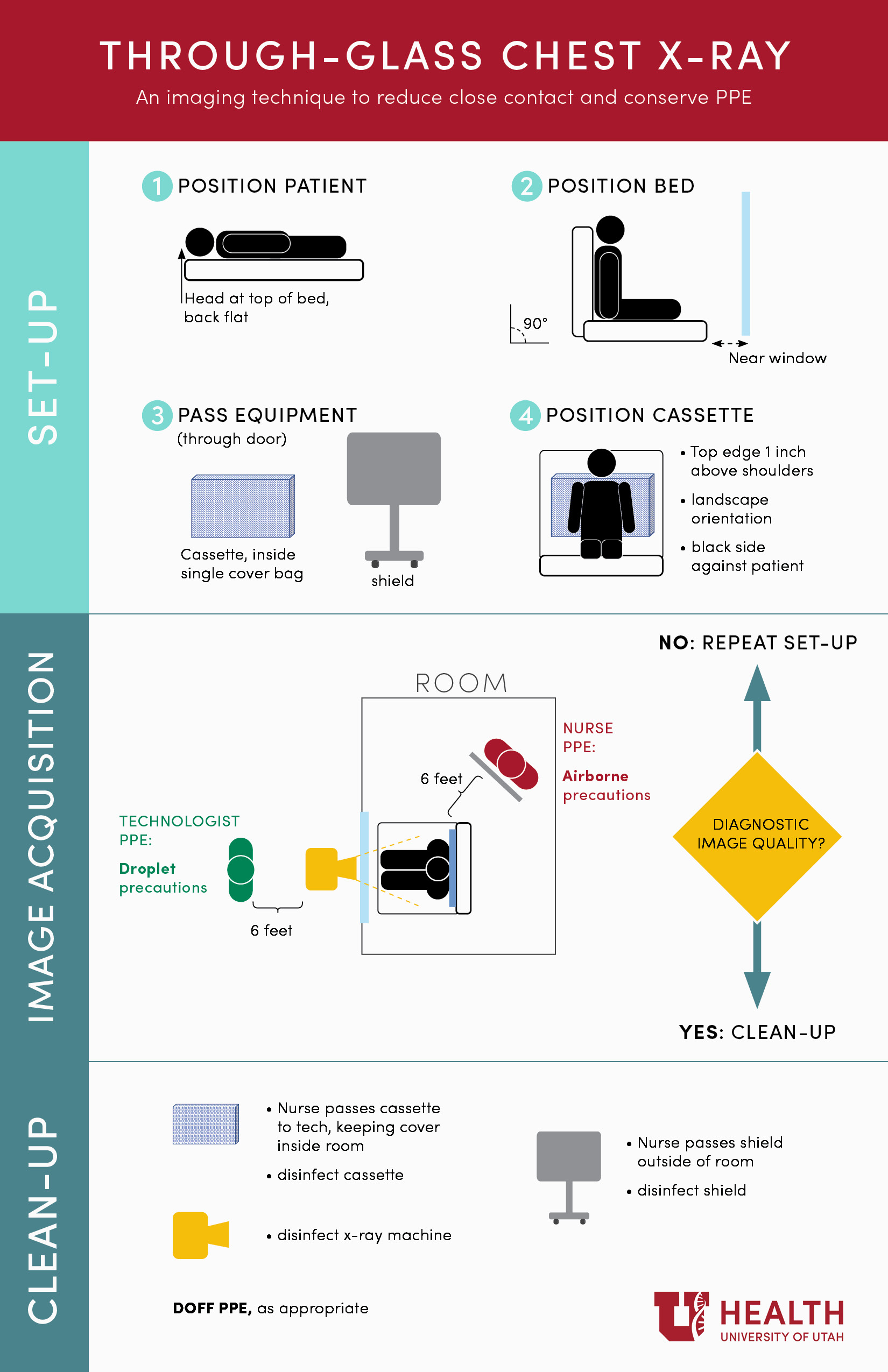
Radiologists and radiologic technologists at University of Utah Health are doing their part to minimize the chance of COVID-19 spreading while at the same time conserving precious Personal Protective Equipment (PPE). They’re using an innovative technique at University Hospital for taking x-rays of patients with respiratory illnesses. In order to keep suspected COVID-19 patients safely in negative pressure observation rooms, they obtain x-ray images with a portable x-ray machine through the glass wall of the room. (scroll to bottom of page for instructional video)
“With some minor technical modifications, chest x-rays taken through the glass are just as clear as normal x-rays,” states Phuong-Anh Duong, MD, Associate Professor of Radiology. Duong, along with her colleagues in the Cardiothoracic Imaging Section, Joyce Schroeder, MD (Section Chief), and Howard Mann, MD, brought the idea to Alex Nieves, MPA, the Manager of Diagnostic Radiology services at the hospital. Together, they tested the new setup and were pleased with the results.
Normally, inpatients are taken to x-ray rooms where technologists position them and obtain the images. Even in the Emergency Department, portable x-ray machines are brought into rooms and patients are positioned by staff so that the images can be obtained correctly. With the advent of the COVID-19 pandemic, University Hospital policy is that any patient who comes to the hospital struggling with respiratory symptoms is treated as a suspected COVID-19 infection. These patients are taken directly to the Emergency Care Unit (separate, negative-pressure rooms from standard ED rooms). The goal is to minimize contact between these patients and the rest of the hospital, including care providers.
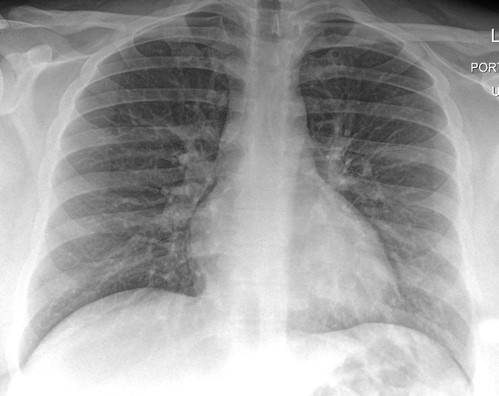
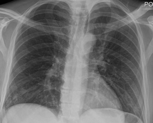
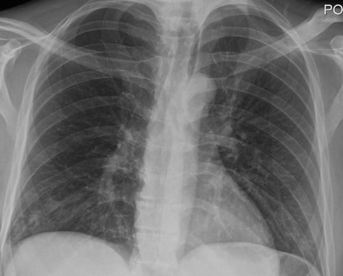
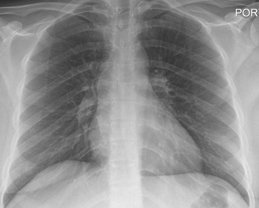
Chest x-rays taken with the new through-the-glass technique.
|
|
|
|
Phuong-Anh Duong, MD |
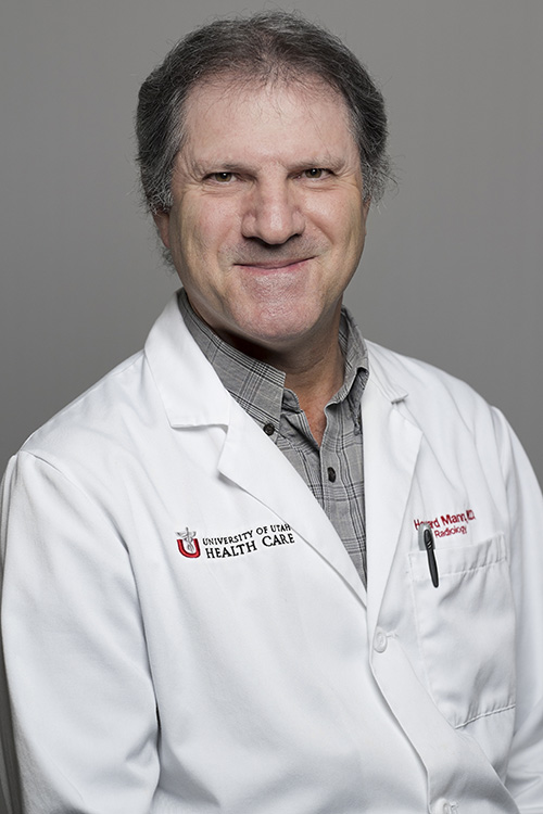
Howard Mann, MD |
|
|
|
|
Joyce Schroeder, MD |
Alex Nieves, MPA |
Taking a chest x-ray is one of the first tests done for patients with respiratory distress. “This technique minimizes hallway traffic for patients and staff, which greatly decreases the risk for transmitting COVID-19,” explains Nieves. What’s more, with concerns over PPE being scarce across the country (although this is not a concern in Utah at the moment), staff do not have to use a set of masks, goggles, and gloves in order to take these x-rays because they do not need to enter the room. This saves time, exposure risk, and cost of materials needed for PPE and sanitization of equipment. “We’re using this technique every day,” notes Nieves.
Dr. Mann was first made aware of this technique from colleagues at the University of Washington Medical Center who began experimenting with it at the end of March. He brought it to our team and we quickly followed suit. We are working on refining the technique, providing training for nurses who will help with the positioning, and studying the effectiveness of this technique in reducing the contact between sick patients and radiology personnel and equipment.
“We are trying to do our part to be as innovative as possible in meeting this ever-changing situation,” notes Satoshi Minoshima, MD, PhD, the Anne G. Osborn Chair of Radiology and Imaging Sciences. “We don’t know how long this situation might last and finding ways to safely treat patients and conserve PPE is paramount,” he adds.
